interna dr. b. rudi utantio - chd_congenital heart disease 2.ppt
TRANSCRIPT
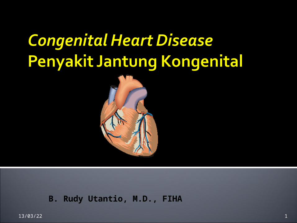
B. Rudy Utantio, M.D., FIHA
19/04/23 1

Tingkat Kemampuan 2 mendiagnosis dan merujukLulusan dokter mampu membuat diagnosis klinik terhadap penyakit tersebut dan menentukan rujukan yang paling tepat bagi penanganan pasien selanjutnya. Lulusan dokter juga mampu menindaklanjuti sesudah kembali dari rujukan
19/04/23 2

Left-to-Right Shunt Lesions
Atrial Septal Defect (ASD)Ventricular Septal Defect (VSD)Atrioventricular Septal Defect (AV
Canal)Patent Ductus Arteriosus (PDA)
19/04/23 3

ASD adalah defek / terbukanya septum interatrial yang menyebabkan terjadinya hubungan yang bebas darah di atrium kiri dengan darah di atrium kanan.
Prevalensi 5-10% PJB
19/04/23 4

ASD Sekundum – di foramen ovale, paling sering didapatkan
• ASD Primum – fusi yang tidak sempurna antara septum primum dengan endocardial cushion, terjadi di bagian bawah foramen ovale dekat katup mitral dan trikuspid yang dapat menyebabkan MR atau TR
• ASD Sinus Venosus – terletak di bagian superior dari foramen ovale dan dekat muara vena kava superior
19/04/23 5

ASD sekundum ASD Sinus venosus
19/04/23 6

Keluhan dan gejala klinik
• Pada umumnya asimptomatik, bisa cepat lelah atau pertumbuhan yang lambat , bila terjadi gagal jantung keluhan utamanya sesak
• Bila terjadi hipertensi pulmonal, bisa nampak sianosis (sindroma Eisenmenger)
19/04/23 7

Pemeriksaan fisik :• Inspeksi : - Prekordium hiperaktif RV heave • Auskultasi : - Suara jantung : fixed widely split S2 - Suara tambahan : systolic ejection
murmur gr II-III/VI @ LSB. Mid-diastolic murmur
terdengar di LLSB.
19/04/23 8

Apa yang menyebabkan bising sistolik dan bising diastolik pada ASD?
• Bising sistolik disebabkan oleh
meningkatnya aliran darah yang melalui katup pulmonal, bukan karena defek pada ASD
Bising diastolik disebabkan karena meningkatnya aliran darah melalui katup trikuspid
19/04/23 9

Medikamentosa
Pengobatan diberikan bila terjadi gagal jantung
Profilaksis terhadap infeksi endokarditis tidak diperlukan kecuali bila ada prolaps katup mitral
19/04/23 10
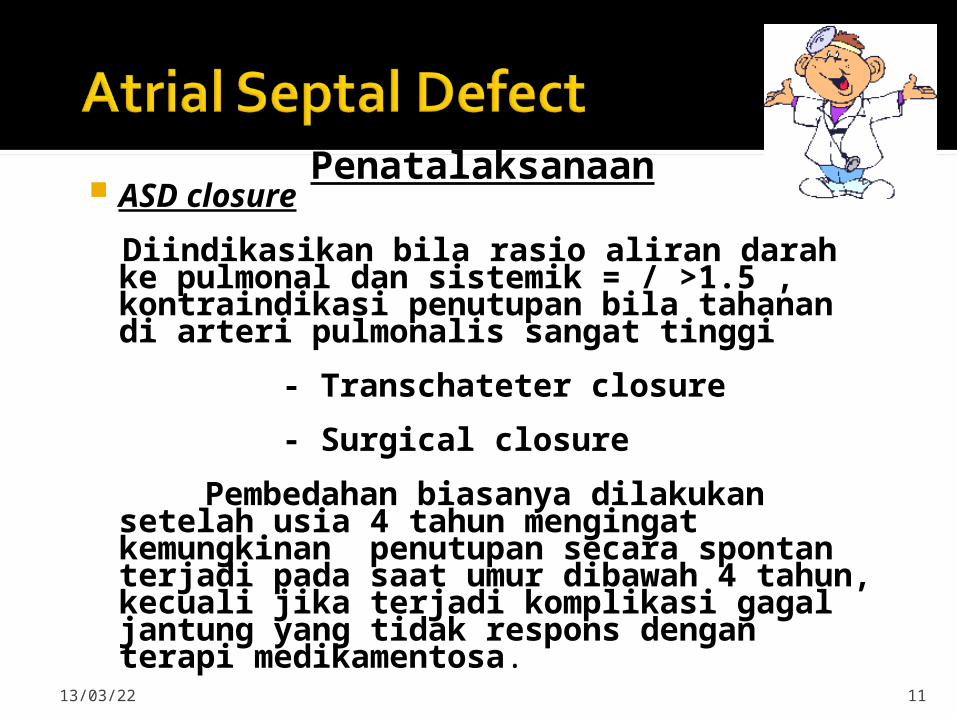
Penatalaksanaan ASD closure
Diindikasikan bila rasio aliran darah ke pulmonal dan sistemik = / >1.5 , kontraindikasi penutupan bila tahanan di arteri pulmonalis sangat tinggi
- Transchateter closure
- Surgical closure
Pembedahan biasanya dilakukan setelah usia 4 tahun mengingat kemungkinan penutupan secara spontan terjadi pada saat umur dibawah 4 tahun, kecuali jika terjadi komplikasi gagal jantung yang tidak respons dengan terapi medikamentosa.
19/04/23 11

19/04/23 12

VSD Defek pada septum interventrikel, bisa
satu atau lebih (swiss cheese VSD) , terjadi akibat kegagalan fusi septum intraventrikular semasa janin.
VSD merupakan PJ kongenital yang paling sering terjadi, 15-20% PJ Kongenital
19/04/23 13

4 Types :
Perimembranous ( membranous) – paling sering.
Infundibular (perimembranous outlet, subpulmonary or supracristal VSD) – mempengaruhi RV outflow tract.
Trabecular VSD
• Muscular VSD – central, marginal atau apical.
19/04/23 14
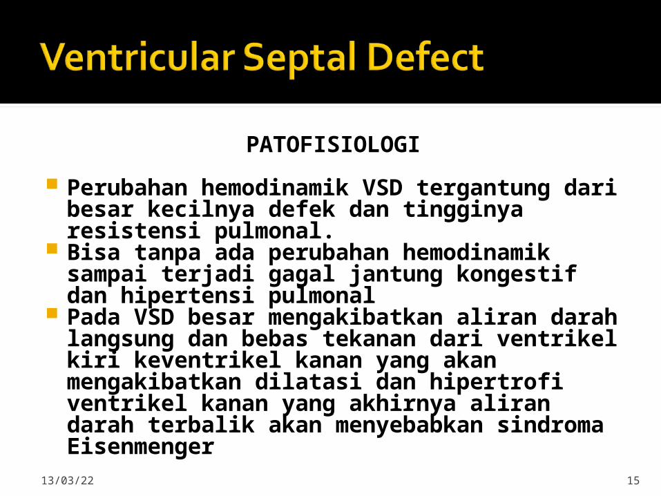
PATOFISIOLOGI
Perubahan hemodinamik VSD tergantung dari besar kecilnya defek dan tingginya resistensi pulmonal.
Bisa tanpa ada perubahan hemodinamik sampai terjadi gagal jantung kongestif dan hipertensi pulmonal
Pada VSD besar mengakibatkan aliran darah langsung dan bebas tekanan dari ventrikel kiri keventrikel kanan yang akan mengakibatkan dilatasi dan hipertrofi ventrikel kanan yang akhirnya aliran darah terbalik akan menyebabkan sindroma Eisenmenger
19/04/23 15

Clinical Signs & Symptoms
• Small - moderate VSD, 3-6mm, are usually asymptomatic and 50% will close
spontaneously by age 2yrs.
• Moderate – large VSD, almost always have symptoms and will require surgical repair.
19/04/23 16

Clinical Signs & Symptoms
• II-III/VI harsh holosystolic murmur heard along the LSB, more prominent with small VSD, maybe absent with a
very Large VSD.
• Prominent P2, Diastolic murmur.
• CHF, FTT, Respiratory infections, exercise intolerance
hyperactive precordium. Symptoms develop between 1 – 6
months
19/04/23 17

Treatment
• Small VSD - no surgical intervention, no physical restrictions, just reassurance and periodic follow-up and endocarditis
prophylaxis.
• Symptomatic VSD - Medical treatment initially with afterload reducers & diuretics.
19/04/23 18

Treatment
Indications for Surgical Closure:
Large VSD w/ medically uncontrolled symptomatology & continued FTT.
Ages 6-12 mo w/ large VSD & Pulm. HTN
Age > 24 mo w/ Qp:Qs ratio > 2:1.
Supracristal VSD of any size, secondary to risk of developing AV insufficiency.
19/04/23 19

AVSD results from incomplete fusion the the endocardial cushions, which help to form the lower portion of the atrial septum, the membranous portion of the ventricular septum and the septal leaflets of the triscupid and mitral valves.
They account for 4% OF ALL CHD.
19/04/23 20

Question: What genetic disease is AVSD more commonly seen in?
• Answer: Down’s Syndrome (Trisomy 21),
Seen in 20-25% of cases.
19/04/23 21

Complete Form Low primum ASD
continuous with a posterior VSD.
Cleft in both septal leaflets of TV/MV.
Results in a large L to R shunt at both levels.
TR/MR, Pulm HTN w/ increase in PVR.
Incomplete Form Any one of the
components may be present.
Most common is primum ASD, cleft in the MV & small VSD.
Hemodynamics are dependent on the lesions.
19/04/23 22

Complete AVSD
19/04/23 23

Clinical Signs & Symptoms Incomplete AVSD maybe
indistinguishable from ASD - usually asymptomatic.
Congestive heart failure in infancy. Recurrent pulmonary infections. Failure to thrive. Exercise intolerance, easy fatigability. Late cyanosis from pulmonary vascular
disease w/ R to L shunt.
19/04/23 24

Clinical Signs & Symptoms Hyperactive precordium Normal or accentuated 1st hrt sound Wide, fixed splitting of S2 Pulmonary systolic ejection murmur
w/thrill Holosystolic murmur @ apex
w/radiation to axilla Mid-diastolic rumbling murmur @ LSB Marked cardiac enlargement on CX-Ray
19/04/23 25

Treatment Surgery is always required.
Treat congestive symptoms. Pulmonary banding maybe required in
premature infants or infants < 5 kg. Correction is done during infancy to
avoid irreversible pulmonary vascular disease.
Mortality low w/incomplete 1-2% & as high as 5% with complete AVSD.
19/04/23 26

PDA – Persistence of the normal fetal vessel that joins the PA to the Aorta.
Normally closes in the 1st wk of life.
Accounts for 10% of all CHD, seen in 10% of other congenital hrt lesions and can often play a critical role in some lesions.
Female : Male ratio of 2:1
Often associated w/ coarctation & VSD.
19/04/23 27
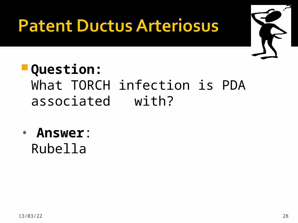
Question:What TORCH infection is PDA associated with?
• Answer: Rubella
19/04/23 28

Hemodynamics As a result of higher aortic pressure,
blood shunts L to R through the ductus from Aorta to PA.
Extent of the shunt depends on size of the ductus & PVR:SVR.
Small PDA, pressures in PA, RV, RA are normal.
19/04/23 29
HemodynamicsLarge PDA, PA pressures are equal to
systemic pressures. In extreme cases 70% of CO is shunted through the ductus to pulmonary circulation.
Leads to increased pulmonary vascular disease.
19/04/23 30

Clinical Signs & Symptoms Small PDA’s are usually asymptomatic Large PDA’s can result in symptoms of
CHF, growth restriction, FTT. Bounding arterial pulses Widened pulse pressure Enlarged heart, prominent apical
impulse Classic continuous machinary systolic
murmur Mid-diastolic murmur at the apex
19/04/23 31
Treatment Indomethacin, inhibitor of prostaglandin
synthesis can be used in premature infants.
PDA requires surgical or catheter closure.
Closure is required treatment heart failure & to prevent pulmonary vascular disease.
Usually done by ligation & division or intra vascular coil.
Mortality is < 1%19/04/23 32

Pulmonary Stenosis
Aortic Stenosis
Coarctation of the Aorta
19/04/23 33

Pulmonary Stenosis is obstruction in the region of either the pulmonary valve or the subpulmonary ventricular outflow tract.
Accounts for 7-10% of all CHD.
Most cases are isolated lesions
Maybe biscuspid or fusion of 2 or more leaflets.
Can present w/or w/o an intact ventricular septum.
19/04/23 34

Question:What syndrome is PS associated with?
Answer:Noonan’s Syndrome, secondary to valve dysplasia.
19/04/23 35
HemodynamicsRV pressure hypertrophy RV
failure.RV pressures maybe > systemic
pressure.Post-stenotic dilation of main PA.W/intact septum & severe stenosis
R-L shunt through PFO cyanosis.Cyanosis is indicative of Critical PS.19/04/23 36

Clinical Signs & Symptoms Depends on the severity of obstruction. Asymptomatic w/ mild PS < 30mmHg. Mod-severe: 30-60mmHg, > 60mmHg Prominent jugular a-wave, RV lift Split 2nd hrt sound w/ a delay Ejection click, followed by systolic
murmur. Heart failure & cyanosis seen in severe
cases.
19/04/23 37
Treatment Mild PS no intervention required, close
follow-up.
Mod-severe – require relieve of stenosis.
Balloon valvuloplasty, treatment of choice.
Surgical valvotomy is also a consideration.
19/04/23 38

Aortic Stenosis is an obstruction to the outflow from the left ventricle at or near the aortic valve that causes a systolic pressure gradient of more than 10mmHg. Accounts for 7% of CHD.
3 Types Valvular – Most common. Subvalvular(subaortic) – involves the left
outflow tract. Supravalvular – involves the ascending
aorta is the least common.
19/04/23 39

Question:Which syndrome is supravalvular stenosis found in?
Answer:Williams Syndrome
19/04/23 40

HemodynamicsPressure hypertrophy of the LV and
LA with obstruction to flow from the LV.
Mild AS 0-25mmHGModerate AS 25-50mmHgSevere AS 50-75mmHgCritical AS > 75mmHg
19/04/23 41

Clinical Signs & Symptoms Mild AS may present with exercise
intolerance, easy fatigabiltity, but usually asymptomatic.
Moderate AS – Chest pain, dypsnea on exertion, dizziness & syncope.
Severe AS – Weak pulses, left sided heart failure, Sudden Death.
19/04/23 42

Clinical Signs & SymptomsLV thrust at the Apex.
Systolic thrill @ rt base/suprasternal notch.
Ejection click, III-IV/VI systolic murmur @ RSB/LSB w/ radiation to the carotids.
19/04/23 43

Treatment Because surgery does not offer a cure it
is reserved for patients with symptoms and a resting gradient of 60-80mmHg.
For subaortic stenosis it is reserved for gradients of 40-50mmHg because of it’s rapidly progressive nature.
Balloon valvuloplasty is the standard of treatment.
19/04/23 44

Treatment Aortic insufficiency & re-stenosis is likely
after surgery and may require valve replacement.
Activity should not be restricted in Mild AS.
Mod-severe AS, no competitive sports.
19/04/23 45
Coarctation- is narrowing of the aorta at varying points anywhere from the transverse arch to the iliac bifurcation.
98% of coarctations are juxtaductal
Male: Female ratio 3:1.
Accounts for 7 % of all CHD.
19/04/23 46

Question:What other heart anomaly is coarctation associated with?
Answer:Bicuspid aortic valve, seen in > 70% of cases.
19/04/23 47

Question:What genetic syndrome is coarctation seen in?
Answer:Turner’s Syndrome
19/04/23 48
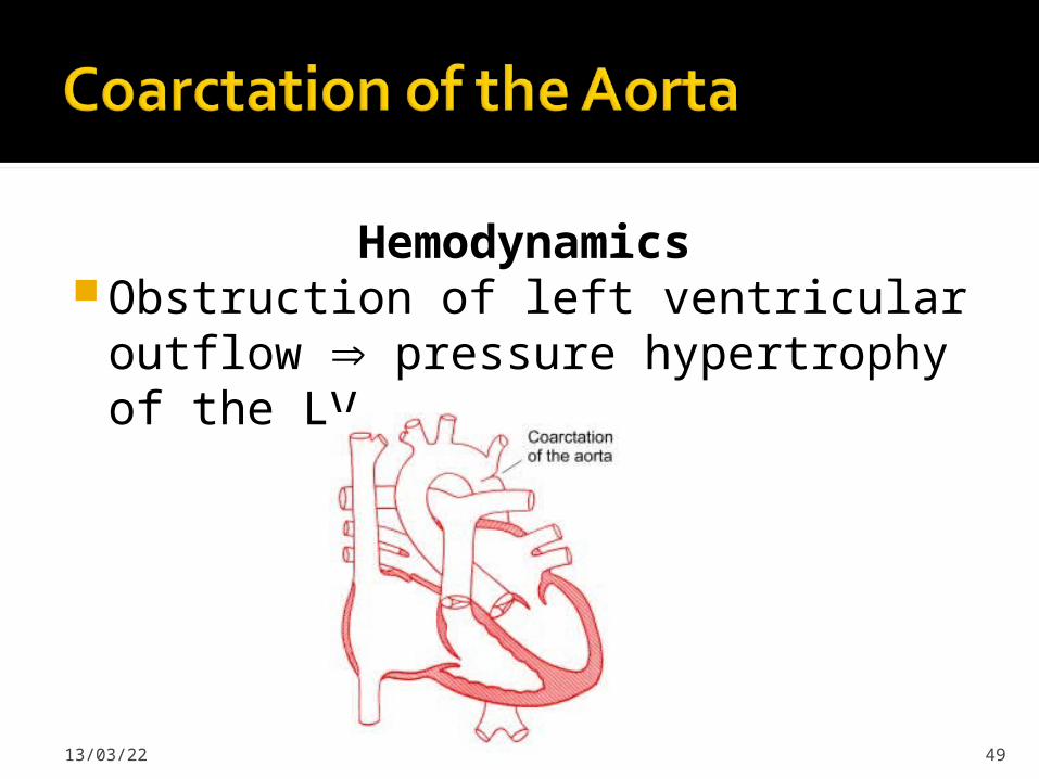
HemodynamicsObstruction of left ventricular
outflow pressure hypertrophy of the LV.
19/04/23 49

Clinical Signs & Symptoms Classic signs of coarctation are
diminution or absence of femoral pulses.
Higher BP in the upper extremities as compared to the lower extremities.
90% have systolic hypertension of the upper extremities.
Pulse discrepancy between rt & lt arms.
19/04/23 50

Clinical Signs & Symptoms With severe coarc. LE hypoperfusion,
acidosis, HF and shock.
Differential cyanosis if ductus is still open
II/VI systolic ejection murmur @ LSB.
Cardiomegaly, rib notching on X-ray.
19/04/23 51

19/04/23 52

Treatment With severe coarctation maintaining the
ductus with prostaglandin E is essential.
Surgical intervention, to prevent LV dysfunction.
Angioplasty is used by some centers.
Re-coarctation can occur, balloon angioplasty is the procedure of choice.
19/04/23 53
Examination of a 3-hr old infant revealsdysmorphic features and cyanosis. Both theocciput and facial profile are flat, and the fontanelle is abnormally enlarged. The space between the great and second toe is wide, andthere is a palmar crease extending across the left palm. Room air oximetry reveals a
saturation 70%.
19/04/23 54

Of the following, the MOST likely lesion to
be found on echocardiography would beA. Atrioventricular septal defectB. Coarctation of the aortaC. Hypoplastic left heartD. Total anomalous pulmonary venous
returnE. Truncus arteriosus
19/04/23 55

After a few days of poor feeding andtachypnea, a 3 week old presents with hypotension, poor central and peripheral pulses, and severe metabolic acidosis. A gallop is audible, and the heart appears enlarged on chest radiography.
Hepatomegaly is marked.
19/04/23 56

Of the following, the BEST intervention to
produce a sustained improvement is A. 100% Oxygen administrationB. Dopamine infusionC. Gamma globulin infusionD. Phenylephrine infusionE. Prostaglandin E infusion
19/04/23 57
A term infant is born with a large ventricular septal
defect. At what age is the infant most likely to first
demonstrate clinical findings of CHFA. 2 daysB. 2 weeksC. 2 monthsD. 6 monthsE. 12 months
19/04/23 58