dbd (1)
DESCRIPTION
pptTRANSCRIPT

DENGUE FEVER/DENGUE HEMORRHAGIC FEVER
Dr. Harun Hudari, SpPD FINASIM

CURRICULUM VITAE
• Nama : Dr. H. Harun Hudari, SpPD, FINASIM• TTL : Palembang, 3 Mei 1970• Pendidikan :
- Dokter FK Universitas Sriwijaya 1996
- Spesialis Penyakit Dalam FK Universitas Sriwijaya 2008• Riwayat Pekerjaan :
1. Dokter PTT Puskesmas Muara Rupit 1997 – 2000
2. Dokter PNS RSUD Dr. MM Dunda Gorontalo 2001 – 2004
3. Dokter Spesialis Penyakit Dalam RSUD Banyuasin
2008 – sekarang
4. Dokter Spesialis Penyakit Dalam FK UNSRI/RSMH
Palembang 2009 – sekarang• HP : 081271621966 / 0711 7301744

About Dengue
• Dengue is one of the most important mosquito-born viral diseases affecting humans.
• Viral life cycle involves humans and the mosquito vector Aedes aegypti. – In the U.S. it has been found that the mosquito Aedes albopictus also
transmits the DEN virus.
• The disease is caused by 4 serotypes of the Dengue virus, a member of the genus Flavivirus: DEN-1, DEN-2, DEN-3, DEN-4.
• Infection with the DEN virus can result in Dengue Fever (DF), Dengue Hemorrhagic Fever (DHF) and Dengue Shock Syndrome (DSS).

Dengue is a tropical febrile disease

992- Chinese Encyclopedia
1780- Philadelphia “break-bone fever”
Benjamin Rush
Dengue Fever has a long history…www.philadelphia-reflections.com/topic/17.htm

Isolated by Albert Sabin in 1944

7

8
Manifestations of the dengue syndrome
• Spectrum of illness:
Asymptomatic
Undifferentiatedfever
No hemorrhage
Unusualhemorrhage
Dengue Fever
DHF 1& 2 DHF 3&4DSS
Dengue Hemorrhagic Fever(plasma leakage)
Symptomatic
Dengue virusInfection

Filoviridae(Ebola, Marburg)
Arenaviridae(Lassa, Junin, Machupo, Guanarito)
Bunyaviridae(CCHF, RVF,
Hantaviruses)
Viral Haemorrhagic Fevers
Flaviviridae(dengue, yellow fever, TBE encephalitides)
EnvelopedRNA viruses

Geographic distribution:
Southeast Asia, the Caribbean,
the Western Pacific Regions.
Other epidemiological features:
High incidence in rainy season;
Spread rapidly;
High mobidity;

Distribution of dengue, Eastern Hemisphere

Distribution of Aedes aegypti (red shaded areas) in the Americas in 1970,and in 1997

13


Dengue Virus
• Family: Flaviviridae• ss +RNA• genome: 10 proteins• 3 Structural
– Coat, deliver RNA to target cells
• 7 Nonstructural• Enveloped• 4 serotypes:
– DEN-1, 2, 3, 4
Goodsell, David. "Dengue Virus." RCSB PDB-101. N.p., n.d. Web. 23 Oct. 2012.

17

18

19


Ag
/Ab
level
Day
IgMIgG
Immune Response
SymptomNS1 AgDA
Y-7 -6 -5 -4 -3 -2 -1 1 2 3 4 5 6 7 8 9
10
11
12
AntibodyBite
NS1 Ag
ACUTEPHASE
CONVALES-ENCE PHASE
CRITICAL PHASE
23

Dengue Fever
Febrile Phase
Critical Phase
Recovery Phase

25
Clinical course of dengue infection -
Febrile Phase
Critical Phase
RecoveryPhase
Lasts for 2 – 7 daysClinical features are indistinguishable between DF and DHF
Happens often after the 3rd day of feverClinical presentation depends on the presence and degree of plasma leakageLasts for about 24-48 hours
In DHF patients – plasma leakage stops and is followed by reabsorption of extravascular fluid


27
Febrile Phase• Usually lasts for 2 – 7 days• Fever is often accompanied by
– Facial flushing, skin erythema, generalised body ache, myalgia, arthalgia and headache
– Anorexia, nausea and vomiting are common• Mild hemorrhagic manifestations may be seen
– This may include positive tourniquet test, petechiae or mucosal bleeding
• Earliest abnormality in FBC is a progressive decrease in total WBC count
• These features are indistinguishable between DF and DHF

28
Critical Phase - 1
• Occurs either– Towards the late febrile phase
• Often after 3rd day of fever
or– Around defervescence
• Usually between 3rd day to 5th day of fever; but may go up to the 7th day of fever.
• This phase lasts for 24- 48 hours

29
Critical Phase - 2During this phase if,
• Minimal or no plasma leakage occurs– Patient feels better as the
temperature subsides
• Dengue fever
• Critical volume of plasma leakage occurs– Patient develops DHF– Varying degrees of
circulatory disturbances occur depending on the degree of plasma leakage

30
Critical Phase -3
• In more severe form of plasma leakage– Patients may sweat, become restless, have cool
extremities and prolonged capillary filling time– The pulse rate increases, diastolic BP increases
and the pulse pressure narrows

12011010090807060
Blood pressure, pulse pressure, heart rate in hypovolemic shock
Time
Compensated shock Decompensated shock

32
Critical Phase - 3Clinical warning signs of severe dengue or
high possibility of rapid progression to shock

33
Critical phase - 4• Thrombocytopenia and hemoconcentration are
usually detectable before the onset of shock• HCT level correlates well with plasma volume
loss and disease severity.• However HCT values may be equivocal and hence
unhelpful when there is frank hemorrhage or with untimely HCT determinations

34
Recovery Phase• Plasma leakage stops after 24-48 hours of
defervescence• This followed by reabsorption of extravascular fluid• Patients’ general well being improves, appetite
returns, gastrointestinal symptoms abate, hemodynamic status stabilises and diuresis ensues.
• Recovery of platelet count is typically preceded by the recovery of WCC count

35
WHO classification - DHF• Grade 1
In the presence of haemoconcentration, fever and symptoms, a positive TT and/or easy bruising
• Grade 2spontaneous bleeding in addition to the manifestation from Grade 1
• Grade 3*circulatory failure – rapid, week pulse and narrowing of pulse pressure or
hypotension with the presence of cold, clammy skin and restlessness.• Grade 4*
profound shock – with undetectable blood pressure or pulse.

36
• Grade 1 & 2 – Non-shock DHF• Grade 3 & 4 – DSS

37
Pathophysiology of DHF - 1• Primary pathophysiological abnormality in DHF
and DSS is an acute increase in vascular permeability
• Plasma leakage results in hemoconcentration and hypovolemia or shock
• Hypovolemia leads to reflex tachycardia and generalised vasoconstriction

38
Clinical manifestations of vasoconstriction in various systems are;– Skin
• coolness, pallor and delayed capillary refill time– Cardiovascular system
• raised diastolic blood pressure and a narrowing pulse pressure – Renal system
• reducing urine output – Gastrointestinal system
• vomiting and abdominal pain– Central nervous system
• lethargy, restlessness, apprehension, reduced level of consciousness– Respiratory system
• tachypnoea (respiratory rate >20/min)
Pathophysiology of DHF - 2

Kasus DBD
1.Demam akut 2-7 hari, bersifat bifasik.
2. Manifestasi perdarahan
• uji tourniquet positif
• petekia, ekimosis, atau purpura
• Perdarahan mukosa, saluran cerna, dan tempat bekas suntikan
• Hematemesis atau melena
3. Trombositopenia < 100.00/pl
4. Kebocoran plasma yang ditandai dengan
•Peningkatan nilai hematrokrit >_ 20 % dari nilai baku sesuai umur dan jenis kelamin.
• Penurunan nilai hematokrit >_ 20 % setelah pemberian cairan yang adekuat
• Efusi pleura, asites, hipoproteinemi39

40

41

42

TATALAKSANA• 1. Demam dengue
Pasien DD dapat berobat jalan, tidak perlu dirawat.
Pada fase demam pasien dianjurkan
• Tirah baring, selama demam.
• Obat antipiretik atau kompres hangat
Asetosal tidak dianjurkan gastritis, perdarahan, /asidosis.
• cairan dan elektrolit per oral, jus buah, sirop, susu, disamping air putih, paling sedikit diberikan selama 2 hari.
• Monitor suhu, jumlah trombosit dan hematokrit sampai fase konvalesen.
43

• 2. DBDJenis Cairan (rekomendasi WHO)
Kristaloid. • Larutan ringer laktat (RL) ,
Larutan ringer asetat (RA), Larutan garam faali (GF)• Dekstrosa 5% dalam larutan ringer laktat (D5/RL)• Dekstrosa 5% dalam larutan ringer asetat (D5/RA)• Dekstrosa 5% dalam 1/2 larutan garam faali (D5/1/2LGF)• (Catatan:Untuk resusitasi syok dipergunakan larutan RL atau
RA tidak boleh larutan yang mengandung dekstran)
Koloid.• Dekstran 40• Plasma• Albumin
44

• Monitoring tanda vital• Koreksi Gangguan Metabolik dan Elektrolit• Pemberian Oksigen• Transfusi Darah
45


Volume Therapy Fluid replacement : renal, enteral or dermal loss (interstitial space)
Crystalloids distributes in the intravasuclar and interstitial space filling thereby the extravascular space up
Volume replacement : acute or subacute loss from the intravasuclar - Colloids contain oncotic substances- Ideally the volume stays intravasal
General Fluid Replacement Crystalloids : Compensation extracellular fluid losses
Plasma Volume Replacement Colloids : Compensation of acute or subacute intravascular fluid losses

Perbedaan Terapi berdasarkan kompartemen cairan
Terapi kasus dehidrasi
penggantian kehilangan cairan di semua ruang extracellular
49
Terapi kasus hypovolemia
penggantian kehilangan
cairan di ruang intravascular (= kehilangan darah)
fluid replacement volume replacement
Kristaloid Koloid

Efek osmolarity cairan pada sel

Saat menderita DBD, cairan tubuh banyak hilang karena:
KEBOCORAN SEL
DEMAM PERDARAHAN
MUAL & MUNTAH
BAHAYA DEHIDRASI PADA PASIEN DBD :• Demam lambat turunnya • Pematangan trombosit terhambat perdarahan sulit
dihentikan• Kekentalan darah meningkat supply oksigen dan
nutrisi ke seluruh tubuh melambat • Tekanan darah tidak stabil• Kekuatan otot berkurang
KENAPA SAAT DBD PERLU CAIRAN?
Cairan tubuh (air + ION) berkurang.POTENSI
DEHIDRASI

Approach to the Management
Groups A• may be sent
home• tolerate
adequate volumes of oral fluids and pass urine at least once every 6 hours
• no warning signs
Groups B• referred for in-
hospital management
• with warning signs, co-existing conditions,
• with certain social circumstances
Groups C• require
emergency treatment and urgent referral
• severe dengue (in critical phase)
Management Decisions

Group A Action Plan
• Encourage intake of ORS, fruit juice and other fluids• Paracetamol and tepid sponge for fever• Advise to come back if with
no clinical improvement severe abdominal pain persistent vomiting cold and clammy extremities,lethargy or irritability or restlessness, bleeding
not passing urine for more than 4–6 hours.
monitor: temperature pattern, volume of fluid intake and losses, urine output, warning signs, signs of plasma leakage and bleeding, haematocrit, and white blood cell and platelet counts

Group B (with warning signs) Action Plan
• reference hematocrit before fluid therapy• isotonic solutions
5–7 ml/kg/hour for 1–2 hours, then reduce to 3–5 ml/kg/hr for 2–4 hours, and then reduce to 2–3 ml/kg/hr or less according to the clinical response
reassess:• haematocrit remains the same or rises only minimally 2–3 ml/kg/hr for
another 2–4 hours • worsening vital signs and rising haematocrit rising 5–10 ml/kg/hour for 1–2
hours

Group B (with warning signs) Action Plan
Give minimum intravenous fluid volume: maintain good perfusion and urine output of about 0.5 ml/kg/hr• Intravenous fluids are usually needed for only 24–48 hours. • Reduce intravenous fluids gradually when the rate of plasma
leakage decreases towards the end of the critical phase.
monitor: • vital signs and peripheral perfusion (1–4 hourly until the patient is
out of the critical phase)• urine output (4–6 hourly)• hematocrit (before and after fluid replacement, then 6–12 hourly) • blood glucose • organ functions (renal profile, liver profile, coagulation profile)

Group B (without warning signs) Action Plan
• Encourage oral fluids
• If not tolerated, start intravenous fluid therapy of 0.9% saline or Ringer’s lactate with or without dextrose at maintenance rate
Patients may be able to take oral fluids after a few hours of intravenous fluid therapy.
• Give the minimum volume required to maintain good perfusion and urine output.
• Intravenous fluids are usually needed only for 24–48 hours.
• Close monitoring

Group C Action Plan
• admit to a hospital with access to intensive care facilities and blood transfusion
• plasma losses should be replaced immediately and rapidly with isotonic crystalloid solution or, in the case of hypotensive shock, colloid solutions
• blood transfusion: with suspected/severe bleeding
• judicious intravenous fluid resuscitation: sole intervention required

Group C Action Plan
Goals of fluid resuscitation:• improving central and peripheral circulation
(decreasing tachycardia, improving BP, warm and pinkextremities, and capillary refill time <2 seconds)
• improving end-organ perfusion– i.e. stable conscious level (more alert or less restless), urine output ≥ 0.5 ml/kg/hour,decreasing metabolic acidosis.


60

61
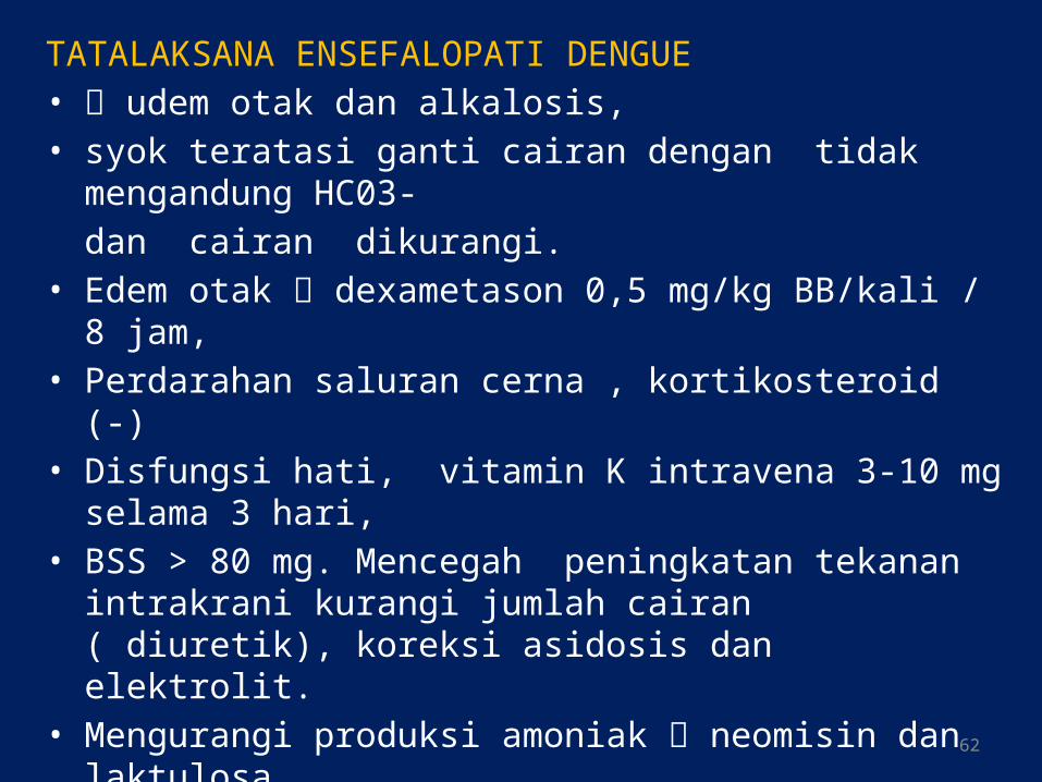
TATALAKSANA ENSEFALOPATI DENGUE• udem otak dan alkalosis,• syok teratasi ganti cairan dengan tidak mengandung HC03-
dan cairan dikurangi.• Edem otak dexametason 0,5 mg/kg BB/kali / 8 jam,• Perdarahan saluran cerna , kortikosteroid (-)• Disfungsi hati, vitamin K intravena 3-10 mg selama 3 hari, • BSS > 80 mg. Mencegah peningkatan tekanan intrakrani kurangi
jumlah cairan ( diuretik), koreksi asidosis dan elektrolit. • Mengurangi produksi amoniak neomisin dan laktulosa. • Transfusi darah • Masa penyembuhan , diberikan asam amino rantai pendek.
62

Kriteria Memulangkan Pasien
memenuhi semua keadaan dibawah ini
1.Tampak perbaikan secara klinis
2.Tidak demam selaina 24 jam tanpa antipiretik
3.Tidak dijumpai distres pernafasan (disebabkan oleh efusi pleura atau asidosis)
4. Hematokrit stabil
5. Jumlah trombosit cenderung naik > 50.000/pl
6. Tiga hari setelah syok teratasi
7. Nafsu makan membaik
63

64
Thank you
Arigatou

Terima kasih
65

66