steroid hipertensi mekanism.pdf
TRANSCRIPT
8/9/2019 STEROID HIPERTENSI MEKANISM.pdf
http://slidepdf.com/reader/full/steroid-hipertensi-mekanismpdf 1/12
81
ORIGINAL PAPER
Nagoya J. Med. Sci. 75. 81 ~ 92, 2013
GLUCOCORTICOID-INDUCED HYPERTENSIONAND CARDIAC INJURY:
EFFECTS OF MINERALOCORTICOID AND
GLUCOCORTICOID RECEPTOR ANTAGONISM
TAKUYA HATTORI1, TAMAYO MURASE1, ERIKA IWASE2, KEIJI TAKAHASHI1,MASAFUMI OHTAKE1, KOJI TSUBOI1, MAYUKO OHTAKE1, MASAAKI MIYACHI1,
TOYOAKI MUROHARA3 and KOHZO NAGATA1
1 Department of Pathophysiological Laboratory Sciences and 3 Department of Cardiology, Nagoya University Graduate School of Medicine, Nagoya, Japan
2 Department of Medical Technology, Nagoya University School of Health Sciences, Nagoya, Japan
ABSTRACT
Glucocorticoids are widely administered for the treatment of various disorders, although their long-term
use results in adverse effects associated with glucocorticoid excess. We investigated the pathophysiological
roles of glucocorticoid receptors (GRs) and mineralocorticoid receptors (MRs) in the cardiac changes
induced by exogenous corticosterone in rats. Corticosterone or vehicle was injected twice daily in rats from
8 to 12 weeks of age. The effects of the GR antagonist RU486, the MR antagonist spironolactone, or both
agents on corticosterone action were also determined. Corticosterone induced hypertension, left ventricular
(LV) fibrosis, and LV diastolic dysfunction. Neither RU486 nor spironolactone affected corticosterone-induced hypertension, whereas spironolactone, but not RU486, attenuated the effects of corticosterone on
LV fibrosis and diastolic function. Corticosterone also increased cardiac oxidative stress and inflammation
in a manner sensitive to spironolactone but not to RU486. The corticosterone-induced LV atrophy was not
affected by either RU486 or spironolactone. Our results implicate MRs in the cardiac fibrosis and diastolic
dysfunction, but not MRs or GRs in the cardiac atrophy, induced by corticosterone. Neither MRs nor GRs
appear to contribute to corticosterone-induced hypertension.
Key Words: glucocorticoids, hypertension, cardiac atrophy, diastolic dysfunction
INTRODUCTION
Glucocorticoids are used widely for the treatment of patients with various conditions including
Received: October 9, 2012; accepted: December 13, 2012
Corresponding author: Kohzo Nagata, MD, PhD
Department of Pathophysiological Laboratory Sciences, Nagoya University Graduate School of Medicine,
1-1-20 Daikominami, Higashi-ku, Nagoya 461-8673, Japan
Tel./Fax: +81-52-719-1546. E-mail: [email protected]
The research described in this paper was presented as a poster that was selected as a winner in the poster
competition for the Co-medical Session at the annual meeting of the Japanese Heart Failure Society in 2009.
Source of Funding: This work was supported by unrestricted research grants from Nippon Boehringer Ingelheim
Co. Ltd. (Tokyo, Japan) and Kyowa Hakko Kirin Co. Ltd. (Tokyo, Japan) as well as by Management Expenses
Grants from the Japanese government to Nagoya University.
8/9/2019 STEROID HIPERTENSI MEKANISM.pdf
http://slidepdf.com/reader/full/steroid-hipertensi-mekanismpdf 2/12
82
Kohzo Nagata et al.
autoimmune and allergic diseases as well as lymphoproliferative disorders.1) Although exogenous
glucocorticoids normally act as potent anti-inflammatory agents, their long-term administration
results in several adverse effects associated with glucocorticoid excess.2) Such glucocorticoid ex-
cess is manifested by a variety of symptoms and signs, including central obesity with moon face,osteoporosis, myopathy, and cardiovascular disorders such as hypertension and atherosclerosis.3)
Cardiovascular complications are among the more important such manifestations for predicting the
morbidity and mortality of individuals with glucocorticoid excess,3) with glucocorticoid treatment
also having been associated with heart failure.4)
Both the glucocorticoid receptor (GR) and the mineralocorticoid receptor (MR) are widely
expressed in the cardiovascular system, including the arterial wall and the myocardium, with
glucocorticoids acting directly to maintain vascular tone and to modify vascular inflammatory,
proliferative, and remodeling responses to injury.5,6) Corticosterone, the endogenous glucocorticoid
of rodents, manifests the same affinity for the MR as does aldosterone and is present in blood at
concentrations two orders of magnitude as great as those of aldosterone.7) Whereas endogenous
glucocorticoids bind both the MR and the GR in vivo, aldosterone binds specifically to the MR.Many pathological states characterized by muscle atrophy are associated with an increase
in circulating glucocorticoid levels, suggesting that glucocorticoids might trigger such atrophy.8)
Indeed, administration of high doses of glucocorticoids induces muscular atrophy in humans and
animals.9,10) Studies have suggested that glucocorticoids inhibit protein synthesis and stimulate
protein degradation in skeletal muscle.11,12) An effect of glucocorticoids on the breakdown of
specific skeletal muscle proteins has been described in some13) but not all14) studies. Administration
of high doses of glucocorticoids to animals induces not only a reduction in muscle mass but
also muscle dysfunction.15) The molecular mechanisms by which glucocorticoid excess induces
cardiovascular injury have remained unclear, however.
We have now investigated the effects of exogenous corticosterone on blood pressure as well
as on cardiac remodeling and function in rats, and we have explored the pathophysiological roles
of GRs and MRs in such effects.
METHODS
Animals and experimental protocols
Male inbred Sprague-Dawley (SD) rats were obtained from Japan SLC (Hamamatsu, Japan)
and were handled in accordance with the guidelines of Nagoya University Graduate School of
Medicine as well as with the Guide for the Care and Use of Laboratory Animals (NIH Publication
no. 85–23, revised 1996). The animals were fed normal laboratory chow containing 0.36% NaCl
after 6 weeks of age, and both the diet and tap water were provided ad libitum throughout the
experimental period. At 8 weeks, the rats were randomly allocated to four groups: (1) the CTC
group (n = 6), in which they were administered corticosterone (Sigma, St. Louis, MO, USA) at a
dose of 20 mg per kilogram of body weight per day; (2) the CTC+RU group (n = 6), in which
they were administered both corticosterone (20 mg/kg per day) and RU486 (Sigma) at 2 mg per
day; (3) the CTC+SPL group (n = 6), in which they were administered both corticosterone (20
mg/kg per day) and spironolactone (Sigma) at 20 mg/kg per day; and (4) the CTC+RU+SPL
group (n = 8), in which they were administered corticosterone plus RU486 plus spironolactone
(at the same doses as in the other groups). Corticosterone was injected s.c. twice a day, RU486
was injected s.c. once a day, and spironolactone was administered orally via a gastric tube
once a day, with all drugs being given to the animals from 8 to 12 weeks of age. The doses
of corticosterone, RU486, and spironolactone were determined on the basis of the results of

8/9/2019 STEROID HIPERTENSI MEKANISM.pdf
http://slidepdf.com/reader/full/steroid-hipertensi-mekanismpdf 3/12
83
GLUCOCORTICOID AND CARDIAC INJURY
previous studies.16-18) Untreated SD rats fed normal laboratory chow after 6 weeks of age remain
normotensive, and such animals administered the vehicle for corticosterone served as age-matched
controls (CNT group, n = 6). At 12 weeks, all animals were anesthetized by i.p. injection of
ketamine (50 mg/kg) and xylazine (10 mg/kg) and were subjected to echocardiographic analysis.The heart was subsequently excised, and left ventricular (LV) tissue was separated for analysis.19)
Corticosterone preparation
Corticosterone was reconstituted from a vial containing 500 mg of lyophilized powder with
1 mL of ethanol followed by 24 mL of polyethylene glycol 400 (Sigma) to yield a suspension
of 20 mg/mL.16)
Blood pressure measurement and echocardiographic analysis
Systolic blood pressure (SBP) and heart rate were measured weekly in conscious animals
by tail-cuff plethysmography (BP-98A; Softron Co., Tokyo, Japan). At 12 weeks of age, rats
were subjected to transthoracic echocardiography as described previously. 20) In brief, M-modeechocardiography was performed with a 12.5-MHz transducer (Aplio SSA-700A; Toshiba Medical
Systems, Tochigi, Japan). LV end-diastolic (LVDd) and end-systolic (LVDs) dimensions as well
as the thickness of the interventricular septum (IVST) and LV posterior wall (LVPWT) were
measured, and fractional shortening (FS) was calculated as: [(LVDd – LVDs)/LVDd] × 100%.
For assessment of Doppler-derived indices of LV function, both LV inflow and outflow velocity
patterns were simultaneously recorded by pulsed-wave Doppler echocardiography. For assessment
of LV diastolic function, we calculated the peak flow velocities at the mitral level during rapid
filling (E) and during atrial contraction (A), the E/A ratio, the isovolumic relaxation time (IRT),
and the deceleration time (DcT). Both the isovolumic contraction time (ICT) and ejection time
(ET) were also determined, and the Tei index, which reflects both LV diastolic and systolic
function, was calculated as follows: Tei index = (ICT+IRT)/ET.18)
Histology and immunohistochemistry
LV tissue was fixed in ice-cold 4% paraformaldehyde for 48 to 72 h, embedded in paraffin,
and processed for histology as described.21) To evaluate macrophage infiltration into the myocar-
dium, we performed immunostaining for the monocyte-macrophage marker CD68 with frozen
sections (thickness, 5 μm) that had been fixed with acetone. Endogenous peroxidase activity was
blocked by exposure of the sections to methanol containing 0.3% hydrogen peroxide. Sections
were incubated at 4°C first overnight with mouse monoclonal antibodies to CD68 (clone ED1,
diluted 1:100; Chemicon, Temecula, CA, USA) and then for 30 min with Histofine Sample Stain
Rat MAX PO (Nichirei Biosciences, Tokyo, Japan). Immune complexes were visualized with
diaminobenzidine and hydrogen peroxide, and the sections were counterstained with hematoxylin.
Image analysis was performed with NIH Scion Image software (Scion, Frederick, MD, USA).
Superoxide production
NADPH-dependent superoxide production by homogenates prepared from freshly frozen LV
tissue was measured with an assay based on lucigenin-enhanced chemiluminescence as described
previously.22) The chemiluminescence signal was sampled every minute for 10 min with a
microplate reader (WALLAC 1420 ARVO MX/Light; Perkin Elmer, Waltham, MA, USA), and
the respective background counts were subtracted from experimental values. Lucigenin chemilu-
minescence was expressed as relative light units per milligram of protein. Superoxide produc-
tion in tissue sections was examined with the use of dihydroethidium (Sigma) as described.23)
Dihydroethidium is rapidly oxidized by superoxide to yield fluorescent ethidium, and the sections

8/9/2019 STEROID HIPERTENSI MEKANISM.pdf
http://slidepdf.com/reader/full/steroid-hipertensi-mekanismpdf 4/12
84
Kohzo Nagata et al.
were examined with a fluorescence microscope equipped with a 585-nm long-pass filter. As a
negative control, we performed staining with dihydroethidium after incubation of sections with
superoxide dismutase (300 U/mL) and confirmed that this procedure abolished the fluorescence
(date not shown). The average of dihydroethidium fluorescence intensity values was calculatedwith the use of NIH Image software (ImageJ).24)
Quantitative RT-PCR analysis
Total RNA was extracted from LV tissue and treated with DNase with the use of a spin-
vacuum isolation kit (Promega, Madison, WI, USA). Complementary DNA was synthesized from
2 μg of total RNA by reverse transcription (RT) with random primers (Invitrogen, Carlsbad,
CA, USA) and MuLV Reverse Transcriptase (Applied Biosystems, Foster City, CA, USA).
Real-time polymerase chain reaction (PCR) analysis was performed as previously described25)
with a Prism 7700 Sequence Detector (Perkin Elmer) and with primers and TaqMan probes
specific for cDNAs encoding insulin-like growth factor–1 (IGF-1: 5′-GACCAAGGGGCTTT-
TACTTCAAC-3′, 5′-CCGGAAGCAACACTCATCCAC-3′, and 5′-CCGTCTGTGGTGCCCTC-CGAATGC-3′ as the forward primer, reverse primer, and TaqMan probe, respectively;
GenBank accession no. NM_001082477), myostatin (5′-GACAGTGAGAGAGAGGCGAATG-3 ′,
5′-TGGCTTCTATTCTGGAGTACCTTG-3′, and 5′-TCTCCACGCACACGCATTACACAGCC-3′,
respectively; NM_019151), FOXO1 (5′-CATCACCAAGGCCATCGAGAG-3′, 5′-CACTCTTCAC-
CATCCACTCGTAG-3′, and 5′-CGGAGAAGAGGCTCACCCTGTCGCA-3′, respectively;
XM_342244), FOXO3 (5′-AGACCAGCCACCTTTTCTTCC-3 ′, 5′-CTGAGCGAGTCCGAAGT-
GAG-3′, and 5′-TCGCACTACGGCAACCAGACACTCCA-3′, respectively; NM_001106395),
atrogin-1 (5′-CCGGCCTTCAAAGGTCTCAC-3 ′, 5′-CGCTCAGCCTCTGCATGATG-3′, and
5′-ACCGACCTGCCTGTGTGCTTACAACTG-3 ′, respectively; NM_133521), muscle ring finger–1
(MuRF-1: 5′-GCCATCCTGGACGAGAAGAAG-3′, 5′-GATCAGGGCCTCGATGAAGTC-3 ′, and
5′-CTCCTCCTGCTCCTGAGTGATCCGCTG-3′, respectively; NM_080903), transforming growth
factor–β1 (TGF-β1),20) connective tissue growth factor (CTGF),22) collagen type I,26) and monocyte
chemoattractant protein–1 (MCP-1).22) Reagents for the detection of human glyceraldehyde-
3-phosphate dehydrogenase (GAPDH) mRNA (Applied Biosystems) were used to quantify rat
GAPDH mRNA as an internal standard.
Statistical analysis
Data are presented as means ± SEM. Differences among groups of rats at 12 weeks of age
were assessed by one-way factorial analysis of variance (ANOVA); if a significant difference
was detected, intergroup comparisons were performed with the Fisher multiple-comparison test.
The time course of SBP was compared among groups by two-way repeated-measures ANOVA.
A P value of <0.05 was considered statistically significant.
RESULTS
Physiological analysis
Body weight of rats was significantly smaller in the CTC, CTC+RU, CTC+SPL, and
CTC+RU+SPL groups than in the CNT group at 12 weeks of age (Table 1). Tibial length
was shorter in CTC rats than in CNT rats, and this effect of corticosterone was prevented by
coadministration of RU486 either alone or together with spironolactone, suggesting that inhibition
of bone growth by corticosterone was mediated via GRs. Whereas CNT rats maintained a normal
SBP throughout the experimental period, CTC rats showed a substantial increase in SBP at 9
8/9/2019 STEROID HIPERTENSI MEKANISM.pdf
http://slidepdf.com/reader/full/steroid-hipertensi-mekanismpdf 5/12
85
GLUCOCORTICOID AND CARDIAC INJURY
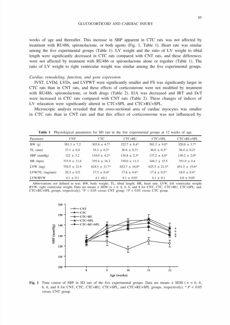
weeks of age and thereafter. This increase in SBP apparent in CTC rats was not affected by
treatment with RU486, spironolactone, or both agents (Fig. 1, Table 1). Heart rate was similar
among the five experimental groups (Table 1). LV weight and the ratio of LV weight to tibial
length were significantly decreased in CTC rats compared with CNT rats, and these differenceswere not affected by treatment with RU486 or spironolactone alone or together (Table 1). The
ratio of LV weight to right ventricular weight was similar among the five experimental groups.
Cardiac remodeling, function, and gene expression
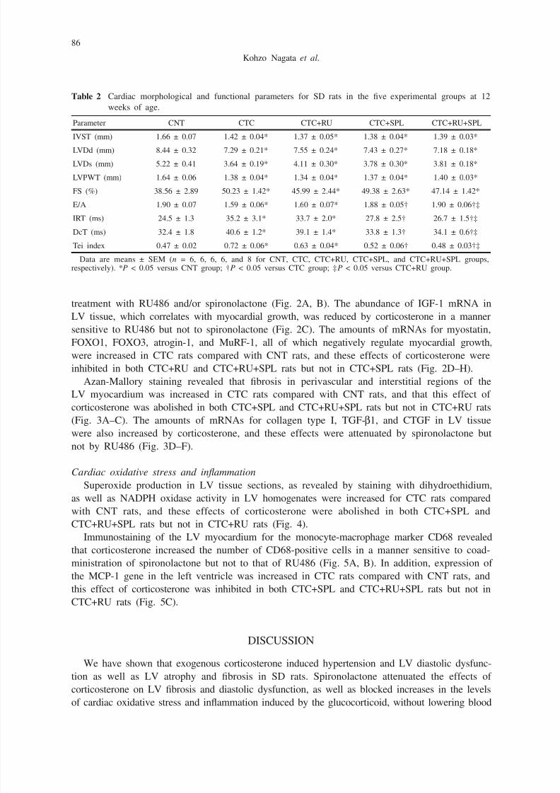
IVST, LVDd, LVDs, and LVPWT were significantly smaller and FS was significantly larger in
CTC rats than in CNT rats, and these effects of corticosterone were not modified by treatment
with RU486, spironolactone, or both drugs (Table 2). E/A was decreased and IRT and DcT
were increased in CTC rats compared with CNT rats (Table 2). These changes of indices of
LV relaxation were significantly altered in CTC+SPL and CTC+RU+SPL.
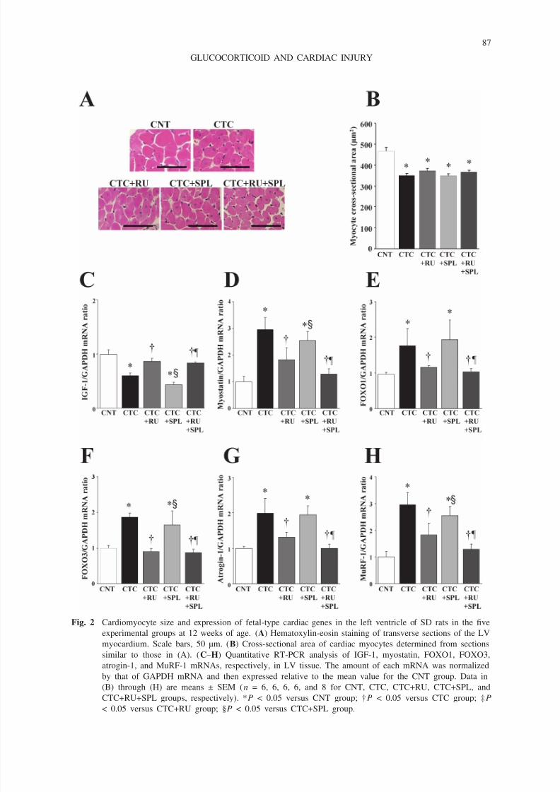
Microscopic analysis revealed that the cross-sectional area of cardiac myocytes was smaller
in CTC rats than in CNT rats and that this effect of corticosterone was not influenced by
Table 1 Physiological parameters for SD rats in the five experimental groups at 12 weeks of age.
Parameter CNT CTC CTC+RU CTC+SPL CTC+RU+SPL
BW (g) 381.3 ± 7.2 303.8 ± 4.7* 322.7 ± 8.4* 301.3 ± 9.0* 320.0 ± 3.7*
TL (mm) 37.1 ± 0.4 35.3 ± 0.3* 36.6 ± 0.3† 36.0 ± 0.3* 36.4 ± 0.2†
SBP (mmHg) 122 ± 3.2 134.0 ± 4.2* 134.8 ± 2.3* 137.2 ± 4.6* 130.2 ± 2.0*
HR (bpm) 333.9 ± 11.6 355.4 ± 16.3 339.0 ± 11.5 348.2 ± 15.5 351.0 ± 8.4
LVW (mg) 754.5 ± 21.9 624.5 ± 11.7* 652.7 ± 18.0* 625.5 ± 21.5* 651.5 ± 15.6*
LVW/TL (mg/mm) 20.3 ± 0.5 17.5 ± 0.4* 17.8 ± 0.4* 17.4 ± 0.5* 18.0 ± 0.4*
LVW/RVW 4.1 ± 0.1 4.1 ±0.1 4.1 ± 0.03 4.1 ± 0.1 4.0 ± 0.05
Abbreviations not defined in text: BW, body weight; TL, tibial length; HR, heart rate; LVW, left ventricular weight;RVW, right ventricular weight. Data are means ± SEM (n = 6, 6, 6, 6, and 8 for CNT, CTC, CTC+RU, CTC+SPL, andCTC+RU+SPL groups, respectively). *P < 0.05 versus CNT group; †P < 0.05 versus CTC group.
Fig. 1 Time course of SBP in SD rats of the five experimental groups. Data are means ± SEM ( n = 6, 6,
6, 6, and 8 for CNT, CTC, CTC+RU, CTC+SPL, and CTC+RU+SPL groups, respectively). *P < 0.05
versus CNT group.
8/9/2019 STEROID HIPERTENSI MEKANISM.pdf
http://slidepdf.com/reader/full/steroid-hipertensi-mekanismpdf 6/12
86
Kohzo Nagata et al.
treatment with RU486 and/or spironolactone (Fig. 2A, B). The abundance of IGF-1 mRNA in
LV tissue, which correlates with myocardial growth, was reduced by corticosterone in a manner
sensitive to RU486 but not to spironolactone (Fig. 2C). The amounts of mRNAs for myostatin,
FOXO1, FOXO3, atrogin-1, and MuRF-1, all of which negatively regulate myocardial growth,
were increased in CTC rats compared with CNT rats, and these effects of corticosterone were
inhibited in both CTC+RU and CTC+RU+SPL rats but not in CTC+SPL rats (Fig. 2D–H).
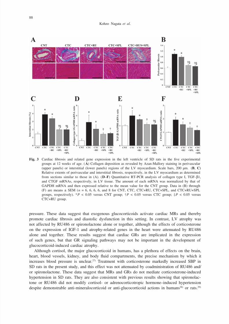
Azan-Mallory staining revealed that fibrosis in perivascular and interstitial regions of the
LV myocardium was increased in CTC rats compared with CNT rats, and that this effect of
corticosterone was abolished in both CTC+SPL and CTC+RU+SPL rats but not in CTC+RU rats
(Fig. 3A–C). The amounts of mRNAs for collagen type I, TGF-β1, and CTGF in LV tissuewere also increased by corticosterone, and these effects were attenuated by spironolactone but
not by RU486 (Fig. 3D–F).
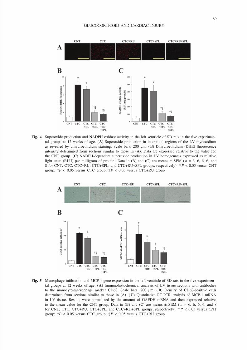
Cardiac oxidative stress and inflammation
Superoxide production in LV tissue sections, as revealed by staining with dihydroethidium,
as well as NADPH oxidase activity in LV homogenates were increased for CTC rats compared
with CNT rats, and these effects of corticosterone were abolished in both CTC+SPL and
CTC+RU+SPL rats but not in CTC+RU rats (Fig. 4).
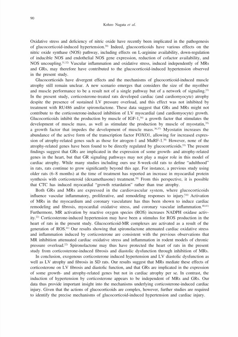
Immunostaining of the LV myocardium for the monocyte-macrophage marker CD68 revealed
that corticosterone increased the number of CD68-positive cells in a manner sensitive to coad-
ministration of spironolactone but not to that of RU486 (Fig. 5A, B). In addition, expression of
the MCP-1 gene in the left ventricle was increased in CTC rats compared with CNT rats, and
this effect of corticosterone was inhibited in both CTC+SPL and CTC+RU+SPL rats but not in
CTC+RU rats (Fig. 5C).
DISCUSSION
We have shown that exogenous corticosterone induced hypertension and LV diastolic dysfunc-
tion as well as LV atrophy and fibrosis in SD rats. Spironolactone attenuated the effects of
corticosterone on LV fibrosis and diastolic dysfunction, as well as blocked increases in the levels
of cardiac oxidative stress and inflammation induced by the glucocorticoid, without lowering blood
Table 2 Cardiac morphological and functional parameters for SD rats in the five experimental groups at 12
weeks of age.
Parameter CNT CTC CTC+RU CTC+SPL CTC+RU+SPL
IVST (mm) 1.66 ± 0.07 1.42 ± 0.04* 1.37 ± 0.05* 1.38 ± 0.04* 1.39 ± 0.03*LVDd (mm) 8.44 ± 0.32 7.29 ± 0.21* 7.55 ± 0.24* 7.43 ± 0.27* 7.18 ± 0.18*
LVDs (mm) 5.22 ± 0.41 3.64 ± 0.19* 4.11 ± 0.30* 3.78 ± 0.30* 3.81 ± 0.18*
LVPWT (mm) 1.64 ± 0.06 1.38 ± 0.04* 1.34 ± 0.04* 1.37 ± 0.04* 1.40 ± 0.03*
FS (%) 38.56 ± 2.89 50.23 ± 1.42* 45.99 ± 2.44* 49.38 ± 2.63* 47.14 ± 1.42*
E/A 1.90 ± 0.07 1.59 ± 0.06* 1.60 ± 0.07* 1.88 ± 0.05† 1.90 ± 0.06†‡
IRT (ms) 24.5 ± 1.3 35.2 ± 3.1* 33.7 ± 2.0* 27.8 ± 2.5† 26.7 ± 1.5†‡
DcT (ms) 32.4 ± 1.8 40.6 ± 1.2* 39.1 ± 1.4* 33.8 ± 1.3† 34.1 ± 0.6†‡
Tei index 0.47 ± 0.02 0.72 ± 0.06* 0.63 ± 0.04* 0.52 ± 0.06† 0.48 ± 0.03†‡
Data are means ± SEM (n = 6, 6, 6, 6, and 8 for CNT, CTC, CTC+RU, CTC+SPL, and CTC+RU+SPL groups,respectively). *P < 0.05 versus CNT group; †P < 0.05 versus CTC group; ‡P < 0.05 versus CTC+RU group.
8/9/2019 STEROID HIPERTENSI MEKANISM.pdf
http://slidepdf.com/reader/full/steroid-hipertensi-mekanismpdf 7/12
87
GLUCOCORTICOID AND CARDIAC INJURY
Fig. 2 Cardiomyocyte size and expression of fetal-type cardiac genes in the left ventricle of SD rats in the five
experimental groups at 12 weeks of age. (A) Hematoxylin-eosin staining of transverse sections of the LV
myocardium. Scale bars, 50 µm. (B) Cross-sectional area of cardiac myocytes determined from sections
similar to those in (A). (C–H) Quantitative RT-PCR analysis of IGF-1, myostatin, FOXO1, FOXO3,
atrogin-1, and MuRF-1 mRNAs, respectively, in LV tissue. The amount of each mRNA was normalized
by that of GAPDH mRNA and then expressed relative to the mean value for the CNT group. Data in
(B) through (H) are means ± SEM (n = 6, 6, 6, 6, and 8 for CNT, CTC, CTC+RU, CTC+SPL, and
CTC+RU+SPL groups, respectively). *P < 0.05 versus CNT group; †P < 0.05 versus CTC group; ‡P
< 0.05 versus CTC+RU group; §P < 0.05 versus CTC+SPL group.
8/9/2019 STEROID HIPERTENSI MEKANISM.pdf
http://slidepdf.com/reader/full/steroid-hipertensi-mekanismpdf 8/12
88
Kohzo Nagata et al.
pressure. These data suggest that exogenous glucocorticoids activate cardiac MRs and thereby
promote cardiac fibrosis and diastolic dysfunction in this setting. In contrast, LV atrophy was
not affected by RU486 or spironolactone alone or together, although the effects of corticosterone
on the expression of IGF-1 and atrophy-related genes in the heart were attenuated by RU486
alone and together. These results suggest that cardiac GRs are implicated in the expression
of such genes, but that GR signaling pathways may not be important in the development of
glucocorticoid-induced cardiac atrophy.
Although cortisol, the major glucocorticoid in humans, has a plethora of effects on the brain,
heart, blood vessels, kidney, and body fluid compartments, the precise mechanism by which it
increases blood pressure is unclear.27) Treatment with corticosterone markedly increased SBP in
SD rats in the present study, and this effect was not attenuated by coadministration of RU486 and/
or spironolactone. These data suggest that MRs and GRs do not mediate corticosterone-induced
hypertension in SD rats. They are also consistent with previous results showing that spironolac-
tone or RU486 did not modify cortisol- or adrenocorticotropic hormone–induced hypertensiondespite demonstrable anti-mineralocorticoid or anti-glucocorticoid actions in humans28) or rats.29)
Fig. 3 Cardiac fibrosis and related gene expression in the left ventricle of SD rats in the five experimental
groups at 12 weeks of age. (A) Collagen deposition as revealed by Azan-Mallory staining in perivascular
(upper panels) or interstitial (lower panels) regions of the LV myocardium. Scale bars, 200 µm. (B, C)
Relative extents of perivascular and interstitial fibrosis, respectively, in the LV myocardium as determined
from sections similar to those in (A). (D–F) Quantitative RT-PCR analysis of collagen type I, TGF-β1,
and CTGF mRNAs, respectively, in LV tissue. The amount of each mRNA was normalized by that of
GAPDH mRNA and then expressed relative to the mean value for the CNT group. Data in (B) through
(F) are means ± SEM (n = 6, 6, 6, 6, and 8 for CNT, CTC, CTC+RU, CTC+SPL, and CTC+RU+SPL
groups, respectively). *P < 0.05 versus CNT group; †P < 0.05 versus CTC group; ‡P < 0.05 versus
CTC+RU group.
8/9/2019 STEROID HIPERTENSI MEKANISM.pdf
http://slidepdf.com/reader/full/steroid-hipertensi-mekanismpdf 9/12
89
GLUCOCORTICOID AND CARDIAC INJURY
Fig. 4 Superoxide production and NADPH oxidase activity in the left ventricle of SD rats in the five experimen-
tal groups at 12 weeks of age. (A) Superoxide production in interstitial regions of the LV myocardium
as revealed by dihydroethidium staining. Scale bars, 200 µm. (B) Dihydroethidium (DHE) fluorescence
intensity determined from sections similar to those in (A). Data are expressed relative to the value for
the CNT group. (C) NADPH-dependent superoxide production in LV homogenates expressed as relative
light units (RLU) per milligram of protein. Data in (B) and (C) are means ± SEM (n = 6, 6, 6, 6, and
8 for CNT, CTC, CTC+RU, CTC+SPL, and CTC+RU+SPL groups, respectively). *P < 0.05 versus CNT
group; †P < 0.05 versus CTC group; ‡P < 0.05 versus CTC+RU group.
Fig. 5 Macrophage infiltration and MCP-1 gene expression in the left ventricle of SD rats in the five experimen-
tal groups at 12 weeks of age. (A) Immunohistochemical analysis of LV tissue sections with antibodies
to the monocyte-macrophage marker CD68. Scale bars, 200 µm. (B) Density of CD68-positive cells
determined from sections similar to those in (A). (C) Quantitative RT-PCR analysis of MCP-1 mRNA
in LV tissue. Results were normalized by the amount of GAPDH mRNA and then expressed relative
to the mean value for the CNT group. Data in (B) and (C) are means ± SEM ( n = 6, 6, 6, 6, and 8
for CNT, CTC, CTC+RU, CTC+SPL, and CTC+RU+SPL groups, respectively). *P < 0.05 versus CNT
group; †P < 0.05 versus CTC group; ‡P < 0.05 versus CTC+RU group.
8/9/2019 STEROID HIPERTENSI MEKANISM.pdf
http://slidepdf.com/reader/full/steroid-hipertensi-mekanismpdf 10/12
90
Kohzo Nagata et al.
Oxidative stress and deficiency of nitric oxide have recently been implicated in the pathogenesis
of glucocorticoid-induced hypertension.30) Indeed, glucocorticoids have various effects on the
nitric oxide synthase (NOS) pathway, including effects on L-arginine availability, down-regulation
of inducible NOS and endothelial NOS gene expression, reduction of cofactor availability, andNOS uncoupling.31,32) Vascular inflammation and oxidative stress, induced independently of MRs
and GRs, may therefore have contributed to the glucocorticoid-induced hypertension observed
in the present study.
Glucocorticoids have divergent effects and the mechanisms of glucocorticoid-induced muscle
atrophy still remain unclear. A new scenario emerges that considers the size of the myofiber
and muscle performance to be a result not of a single pathway but of a network of signaling.33)
In the present study, corticosterone-treated rats developed cardiac (and cardiomyocyte) atrophy
despite the presence of sustained LV pressure overload, and this effect was not inhibited by
treatment with RU486 and/or spironolactone. These data suggest that GRs and MRs might not
contribute to the corticosterone-induced inhibition of LV myocardial (and cardiomyocyte) growth.
Glucocorticoids inhibit the production by muscle of IGF-1,34) a growth factor that stimulates thedevelopment of muscle mass, as well as stimulate the production by muscle of myostatin,35)
a growth factor that impedes the development of muscle mass. 36,37) Myostatin increases the
abundance of the active form of the transcription factor FOXO1, allowing for increased expres-
sion of atrophy-related genes such as those for atrogen-1 and MuRF-1.38) However, none of the
atrophy-related genes have been found to be directly regulated by glucocorticoids.33) The present
findings suggest that GRs are implicated in the expression of some growth- and atrophy-related
genes in the heart, but that GR signaling pathways may not play a major role in this model of
cardiac atrophy. While many studies including ours use 8-week-old rats to define “adulthood”
in rats, rats continue to grow significantly beyond this age. For instance, a previous study using
older rats (6–8 months) at the time of treatment has reported an increase in myocardial protein
synthesis with corticosteroid (dexamethasone) treatment.39) From this perspective, it is possible
that CTC has induced myocardial “growth retardation” rather than true atrophy.
Both GRs and MRs are expressed in the cardiovascular system, where glucocorticoids
influence vascular inflammatory, proliferative, and remodeling responses to injury.5,6) Activation
of MRs in the myocardium and coronary vasculature has thus been shown to induce cardiac
remodeling and fibrosis, myocardial oxidative stress, and coronary vascular inflammation.40,41)
Furthermore, MR activation by reactive oxygen species (ROS) increases NADPH oxidase activ-
ity.22) Corticosterone-induced hypertension may have been a stimulus for ROS production in the
heart of rats in the present study. Glucocorticoid-MR complexes are activated as a result of the
generation of ROS.41) Our results showing that spironolactone attenuated cardiac oxidative stress
and inflammation induced by corticosterone are consistent with the previous observations that
MR inhibition attenuated cardiac oxidative stress and inflammation in rodent models of chronic
pressure overload.22) Spironolactone may thus have protected the heart of rats in the present
study from corticosterone-induced fibrosis and diastolic dysfunction through inhibition of MRs.
In conclusion, exogenous corticosterone induced hypertension and LV diastolic dysfunction as
well as LV atrophy and fibrosis in SD rats. Our results suggest that MRs mediate these effects of
corticosterone on LV fibrosis and diastolic function, and that GRs are implicated in the expression
of some growth- and atrophy-related genes but not in cardiac atrophy per se. In contrast, the
induction of hypertension by corticosterone appears to be independent of MRs and GRs. Our
data thus provide important insight into the mechanisms underlying corticosterone-induced cardiac
injury. Given that the actions of glucocorticoids are complex, however, further studies are required
to identify the precise mechanisms of glucocorticoid-induced hypertension and cardiac injury.
8/9/2019 STEROID HIPERTENSI MEKANISM.pdf
http://slidepdf.com/reader/full/steroid-hipertensi-mekanismpdf 11/12
91
GLUCOCORTICOID AND CARDIAC INJURY
DISCLOSURES
None.
ACKNOWLEDGEMENTS
We thank Masafumi Sakai for technical assistance.
REFERENCES
1) Iuchi T, Akaike M, Mitsui T, Ohshima Y, Shintani Y, Azuma H, Matsumoto T. Glucocorticoid excess induces
superoxide production in vascular endothelial cells and elicits vascular endothelial dysfunction. Circ Res.
2003; 92: 81–87.
2) Saruta T. Mechanism of glucocorticoid-induced hypertension. Hypertens Res. 1996; 19: 1–8.
3) Ross EJ, Linch DC. Cushing’s syndrome--killing disease: discriminatory value of signs and symptoms aiding
early diagnosis. Lancet . 1982; 2: 646–649.
4) Souverein PC, Berard A, Van Staa TP, Cooper C, Egberts AC, Leufkens HG, Walker BR. Use of oral
glucocorticoids and risk of cardiovascular and cerebrovascular disease in a population based case-control
study. Heart . 2004; 90: 859–865.
5) Ullian ME. The role of corticosteriods in the regulation of vascular tone. Cardiovasc Res. 1999; 41: 55–64.
6) Hadoke PW, Macdonald L, Logie JJ, Small GR, Dover AR, Walker BR. Intra-vascular glucocorticoid
metabolism as a modulator of vascular structure and function. Cell Mol Life Sci. 2006; 63: 565–578.
7) Frey FJ, Odermatt A, Frey BM. Glucocorticoid-mediated mineralocorticoid receptor activation and hyperten-
sion. Current opinion in nephrology and hypertension. 2004; 13: 451–458.
8) Schakman O, Gilson H, Kalista S, Thissen JP. Mechanisms of muscle atrophy induced by glucocorticoids.
Horm Res. 2009; 72 Suppl 1: 36–41.
9) Dardevet D, Sornet C, Taillandier D, Savary I, Attaix D, Grizard J. Sensitivity and protein turnoverresponse to glucocorticoids are different in skeletal muscle from adult and old rats. Lack of regulation of
the ubiquitin-proteasome proteolytic pathway in aging. J Clin Invest . 1995; 96: 2113–2119.
10) Mitch WE, Goldberg AL. Mechanisms of muscle wasting. The role of the ubiquitin-proteasome pathway.
N Engl J Med . 1996; 335: 1897–1905.
11) Pereira RM, Freire de Carvalho J. Glucocorticoid-induced myopathy. Joint Bone Spine. 2011; 78: 41–44.
12) Shah OJ, Anthony JC, Kimball SR, Jefferson LS. Glucocorticoids oppose translational control by leucine
in skeletal muscle. Am J Physiol Endocrinol Metab. 2000; 279: E1185–1190.
13) Schakman O, Gilson H, Thissen JP. Mechanisms of glucocorticoid-induced myopathy. J Endocrinol. 2008;
197: 1–10.
14) Odedra BR, Millward DJ. Effect of corticosterone treatment on muscle protein turnover in adrenalectomized
rats and diabetic rats maintained on insulin. Biochem J . 1982; 204: 663–672.
15) Shin YS, Fink H, Khiroya R, Ibebunjo C, Martyn J. Prednisolone-induced muscle dysfunction is caused
more by atrophy than by altered acetylcholine receptor expression. Anesth Analg. 2000; 91: 322–328.16) Mangos GJ, Turner SW, Fraser TB, Whitworth JA. The role of corticosterone in corticotrophin (ACTH)-
induced hypertension in the rat. J Hypertens. 2000; 18: 1849–1855.
17) Robert V, Heymes C, Silvestre JS, Sabri A, Swynghedauw B, Delcayre C. Angiotensin AT1 receptor subtype
as a cardiac target of aldosterone: role in aldosterone-salt-induced fibrosis. Hypertension. 1999; 33: 981–986.
18) Tei C, Ling LH, Hodge DO, Bailey KR, Oh JK, Rodeheffer RJ, Tajik AJ, Seward JB. New index of
combined systolic and diastolic myocardial performance: a simple and reproducible measure of cardiac
function--a study in normals and dilated cardiomyopathy. J Cardiol. 1995; 26: 357–366.
19) Yamada Y, Tsuboi K, Hattori T, Murase T, Ohtake M, Furukawa M, Ueyama J, Nishiyama A, Murohara T,
Nagata K. Mechanism underlying the efficacy of combination therapy with losartan and hydrochlorothiazide
in rats with salt-sensitive hypertension. Hypertens Res. 2011; 34: 809–816.
20) Nagata K, Somura F, Obata K, Odashima M, Izawa H, Ichihara S, Nagasaka T, Iwase M, Yamada Y,
Nakashima N, Yokota M. AT1 receptor blockade reduces cardiac calcineurin activity in hypertensive rats.
Hypertension. 2002; 40: 168–174.21) Miyachi M, Yazawa H, Furukawa M, Tsuboi K, Ohtake M, Nishizawa T, Hashimoto K, Yokoi T, Kojima
8/9/2019 STEROID HIPERTENSI MEKANISM.pdf
http://slidepdf.com/reader/full/steroid-hipertensi-mekanismpdf 12/12
92
Kohzo Nagata et al.
T, Murate T, Yokota M, Murohara T, Koike Y, Nagata K. Exercise training alters left ventricular geometry
and attenuates heart failure in dahl salt-sensitive hypertensive rats. Hypertension. 2009; 53: 701–707.
22) Nagata K, Obata K, Xu J, Ichihara S, Noda A, Kimata H, Kato T, Izawa H, Murohara T, Yokota M. Min-
eralocorticoid receptor antagonism attenuates cardiac hypertrophy and failure in low-aldosterone hypertensive
rats. Hypertension. 2006; 47: 656–664.23) Elmarakby AA, Loomis ED, Pollock JS, Pollock DM. NADPH oxidase inhibition attenuates oxidative stress
but not hypertension produced by chronic ET-1. Hypertension. 2005; 45: 283–287.
24) Murase T, Hattori T, Ohtake M, Nakashima C, Takatsu M, Murohara T, Nagata K. Effects of estrogen on
cardiovascular injury in ovariectomized female DahlS.Z-Lepr(fa)/Lepr(fa) rats as a new animal model of
metabolic syndrome. Hypertension. 2012; 59: 694–704.
25) Somura F, Izawa H, Iwase M, Takeichi Y, Ishiki R, Nishizawa T, Noda A, Nagata K, Yamada Y, Yokota M.
Reduced myocardial sarcoplasmic reticulum Ca(2+)-ATPase mRNA expression and biphasic force-frequency
relations in patients with hypertrophic cardiomyopathy. Circulation. 2001; 104: 658–663.
26) Sakata Y, Yamamoto K, Mano T, Nishikawa N, Yoshida J, Hori M, Miwa T, Masuyama T. Activation of
matrix metalloproteinases precedes left ventricular remodeling in hypertensive heart failure rats: its inhibition
as a primary effect of Angiotensin-converting enzyme inhibitor. Circulation. 2004; 109: 2143–2149.
27) Whitworth JA, Mangos GJ, Kelly JJ. Cushing, cortisol, and cardiovascular disease. Hypertension. 2000; 36:
912–916.28) Clore JN, Estep H, Ross-Clunis H, Watlington CO. Adrenocorticotropin and cortisol-induced changes in
urinary sodium and potassium excretion in man: effects of spironolactone and RU486. J Clin Endocrinol
Metab. 1988; 67: 824–831.
29) Li M, Wen C, Fraser T, Whitworth JA. Adrenocorticotrophin-induced hypertension: effects of mineralocor-
ticoid and glucocorticoid receptor antagonism. J Hypertens. 1999; 17: 419–426.
30) Ong SL, Zhang Y, Whitworth JA. Reactive oxygen species and glucocorticoid-induced hypertension. Clin
Exp Pharmacol Physiol. 2008; 35: 477–482.
31) Radomski MW, Palmer RM, Moncada S. Glucocorticoids inhibit the expression of an inducible, but not
the constitutive, nitric oxide synthase in vascular endothelial cells. Proc Natl Acad Sci U S A. 1990; 87:
10043–10047.
32) Wallerath T, Witte K, Schafer SC, Schwarz PM, Prellwitz W, Wohlfart P, Kleinert H, Lehr HA, Lemmer
B, Forstermann U. Down-regulation of the expression of endothelial NO synthase is likely to contribute to
glucocorticoid-mediated hypertension. Proc Natl Acad Sci U S A. 1999; 96: 13357–13362.
33) Sandri M. Signaling in muscle atrophy and hypertrophy. Physiology (Bethesda). 2008; 23: 160–170.
34) Gayan-Ramirez G, Vanderhoydonc F, Verhoeven G, Decramer M. Acute treatment with corticosteroids
decreases IGF-1 and IGF-2 expression in the rat diaphragm and gastrocnemius. Am J Respir Crit Care
Med . 1999; 159: 283–289.
35) Ma K, Mallidis C, Bhasin S, Mahabadi V, Artaza J, Gonzalez-Cadavid N, Arias J, Salehian B. Glucocor-
ticoid-induced skeletal muscle atrophy is associated with upregulation of myostatin gene expression. Am J
Physiol Endocrinol Metab. 2003; 285: E363–371.
36) McCroskery S, Thomas M, Maxwell L, Sharma M, Kambadur R. Myostatin negatively regulates satellite
cell activation and self-renewal. J Cell Biol. 2003; 162: 1135–1147.
37) Welle S, Bhatt K, Pinkert CA. Myofibrillar protein synthesis in myostatin-deficient mice. Am J Physiol
Endocrinol Metab. 2006; 290: E409–415.
38) McFarlane C, Plummer E, Thomas M, Hennebry A, Ashby M, Ling N, Smith H, Sharma M, Kambadur R.
Myostatin induces cachexia by activating the ubiquitin proteolytic system through an NF-kappaB-independent,
FoxO1-dependent mechanism. J Cell Physiol. 2006; 209: 501–514.
39) Savary I, Debras E, Dardevet D, Sornet C, Capitan P, Prugnaud J, Mirand PP, Grizard J. Effect of gluco-
corticoid excess on skeletal muscle and heart protein synthesis in adult and old rats. The British journal
of nutrition. 1998; 79: 297–304.
40) Nagase M, Fujita T. Mineralocorticoid receptor activation in obesity hypertension. Hypertens Res. 2009;
32: 649–657.
41) Funder JW. RALES, EPHESUS and redox. J Steroid Biochem Mol Biol. 2005; 93: 121–125.