mandibulofacial dysostosis*
TRANSCRIPT
MANDIBULOFACIAL DYSOSTOSIS*
W I T H THE REF
GUILLERMI Santurce,
Mandibulofacial dysostosis (Franceschet-ti's syndrome) is still an extremely uncommon condition but it is now realized that, like all so-called rare diseases, it is more common than it was once supposed to be. The considerable interest recently displayed in it seems to warrant a report of two additional cases, together with a review of the information now available concerning it and the syndromes apparently related to it.
HISTORICAL NOTE
The first reports of mandibulofacial dysostosis are credited to Berry,1 in 1889. His patients, a mother and daughter, presented congenital notches of the lower eyelid, the mother bilaterally and the daughter on the right side only, associated with receding chins.
In 1900, Treacher Collins2 described similar anomalies of the eyelids associated with defects of the malar bones. A number of other reports followed, but it was not until the comprehensive studies of Franceschetti and his associates were published in 19443
and 19494 that mandibulofacial dysostosis was clearly understood. When all the published reports were put together, it was evident that while the associated malformations may vary in degree and extent, mandibulofacial dysostosis is a single clinical entity.
The term Treacher Collins' syndrome was used for this condition after his description of it in 1900, but, from what has just been said, it is obvious that if an eponym is to be used for the disease, the term Franceschetti's syndrome is preferable.
* From the Section of Ophthalmology, University of Puerto Rico School of Medicine. Presented in part at the VI. Pan-American Congress of Ophthalmology, Carcas, Venezuela.
3RT OF TWO CASES
> Pico, M.D. Puerto Rico
CLINICAL CLASSIFICATION
Franceschetti and Klein,4 while pointing out that a sharp distinction cannot be drawn between the categories, divided mandibulofacial dysostosis into five groups, according to the degree of deformity, as follows:
1. The complete form. In the fully developed syndrome the typical findings include:
A. Obliquity of the palpebral fissures downward and laterally (antimongoloid), with a coloboma in the outer portion of the lower lids and sometimes in the upper lids also. The lower lids are S-shaped. The deformity of the palpebral aperture and of the lower lids is due to underdevelopment of the malar bones.
B. Hypoplasia of the facial bones, especially of the malar bones and the mandible, which produces the characteristic fishlike physiognomy with receding chin.
C. Malformation of the external ear and, in some instances, of the middle and inner ear also.
D. Macrostomia, high palate, and abnormal position of the teeth, with resulting maloc-clusion.
E. Blind fistula between the angles of the mouth and the ears.
F. Tongue-shaped growths of hair projecting from the hair line toward the cheek.
G. Other associated anomalies, such as facial clefts and skeletal deformities.
2. The incomplete form. In this form the appearance of the palpebral fissure and the lower lids is characteristic, and there is underdevelopment of the malar bones and the mandible. The external ear may be normal, but hearing is often impaired. In this group, to which Treacher Collins'2 case probably belongs, the syndrome is present but the de-
522 GUILLERMO PICO
velopment is less extensive and less striking. 3. The abortive form. In this group only
the anomalies of the eyelids are present. These abortive cases, according to Duke-Elder,5 are rare. He could find only three cases in the literature, reported, respectively, by Barry in 1889, by Isakowitz in 1927, and by Schachter in 1947. Franceschetti4 commented that it is difficult to decide whether some abortive cases represent a pathologic stigma, the so-called forme fruste of the mandibulof acial syndrome, or should be classified in the "borderline of normality."
4. The unilateral form. In this group the abnormalities, whatever the degree, are present on only one side of the face.
5. The atypical forms. This group includes incomplete forms of the syndrome, in which one of the principal characteristics is missing while other abnormalities, such as microph-thalmos, which do not belong in mandibulo-facial dysostosis, may be present. According to Franceschetti and Klein,4 atypical cases may "lead over" to transitional forms which do not show anomalies of the lids and other typical characteristics and which therefore cannot be classified as mandibulofacial dysostosis.
In a number of the reported cases of this syndrome associated anomalies have been described, including distichiasis, dermolipomas of the conjunctiva, skeletal deformities, and such anomalies of the extremities as syn-dactyly.
PATHOGENESIS
Ida Mann,6 in a report of a case of mandibulofacial dysostosis in 1943, discussed in detail the embryologic meaning of this defect. It appears to originate in a defective gene, which results in defective ossification of the bones of the face that are derived from the maxillary (visceral) mesodermal process of the first visceral (mandibular) arch. Probably the inhibitory factor becomes effective about the seventh week of fetal life, when the facial bones are being formed.
Franceschetti and Klein4 gave a detailed
description of how the various malformations typical of the syndrome appear as a result of the maldevelopment of the facial bones, with resulting hypoplasia and malposition of the surrounding soft tissues of the lips, face, and ears. The blind fistula characteristic of the syndrome is explained as the result of a noncoalescence of the first visceral groove. It is chiefly the mandible and malar bones, and, to a lesser degree, the other facial bones that show defective development. Hypoplasia of the malar bones is the principal cause of the antimongoloid obliquity of the palpebral fissure, as well as of the downward droop of the lateral portion of the eyelids, especially the lower lids, and the S-shape of the border of the lower lids. Colobomas of the lids and occasional associated defects of the lashes and the meibomian glands are secondary, in France-schetti's opinion, to the bony hypoplasia.
HEREDITARY FACTOR
There is no doubt of the hereditary factor in mandibulofacial dysostosis. Several of the genealogies described in the literature indicate that this syndrome is an independent genotypic entity which follows the irregular dominant form of transmission. It was transmitted in uncomplicated form, without associated anomalies, through three generations in the cases observed by Debusmann7 in 1940, Leopold, Mahoney and Price8 in 1945, and Brohm and Kluska9 in 1947.
The marked variability in the degree of the manifestations observed in the reported cases of mandibulofacial dysostosis, which makes it necessary to divide them into a number of different categories, points to an unstable gene, with variable expression. The degree of penetrance of the defective gene varies in the reported cases from the low-grade penetrance of Debusmann's7 cases to the high-grade penetrance in the cases reported by Leopold and his associates.8
Debusmann's report indicates that the gene may have a lethal effect which increases in severity in successive generations. In the
MANDIBULOFACIAL DYSOSTOSIS 523
family that he described, the three children in the last generation, who each presented a fully developed syndrome, died within the first six months of life. The reports of accessory abnormalities, such as anomalies of the vertebral column, the extremities and the thorax, suggest that one is dealing with a pleiotropic (polyphenic) gene.
ALLIED SYNDROMES
Mandibulofacial dysostosis must be differentiated from two other syndromes, the craniofacial dysostosis of Crouzon and the acrocephalosyndactyly of Apert. In both of these conditions, antimongoloid obliquity of the palpebral fissure and S-shaped lower lids may be present, but the appearance of the head and the facial details are in sharp contrast to the fades of the patient with mandib-ulofacial dysostosis.
FAMILIAL PRIMARY HYPOPLASIA OF THE ORBITAL MARGIN
A new syndrome, described in 1955 by Urrets-Zavalia10 and termed familial primary hypoplasia of the orbital margin, also must be considered in this connection. Its principal features are:
1. Marked agenesis of the orbital margin. 2. Hypoplasia of the palpebral skin and
tarsal plates. 3. A variable developmental defect of the
lacrimal passages. In one of the genealogies reported by
Urrets-Zavalia, the defect consisted of a very short inferior canaliculus; ectopia and elongation of the lower punctum; and atresia of the nasolacrimal duct, with a supernumerary canaliculus that opened at the inner canthus. In another genealogy there was a total absence of the lower canaliculus, in conjunction with a shallow, scarcely noticeable colobomatous indentation of the lacrimal portion of the border of the lower lid. A variable amount of hyperopia was frequently present, as well as certain congenital defects of the extraocular muscles.
Urrets-Zavalia explained this malforma
tion as due to a faulty development of the paraxial and visceral mesoderm, which differentiates to form the bones of the upper portion of the face. The abnormalities of the surrounding soft tissues he considered secondary. Hyperopia and insufficiency of some of the extraocular muscles may also be explained by a defective differentiation of the paraxial mesoderm.
The pedigrees of the two affected families showed a dominant type of heredity. The gene demonstrated a highly penetrating character and fairly constant expression. Urrets-Zavalia considered the mode of action of this gene similar to that of the genes held responsible for Crouzon's craniofacial dysostosis; for mandibulofacial dysostosis; and for the deep skeletal and muscular deformities frequently found in association with developmental defects of the external ear. In mandibulofacial dysostosis, Urrets-Zavalia pointed out, the paraxial mesoderm is usually spared, as are also the structures derived from the lateral frontonasal process. For this reason, there is no agenesis of the upper and mesial wall of the orbit in this syndrome, nor are anomalies of the lacrimal ducts usually present.
In Urrets-Zavalia's cases the presence of hypoplasis of the tarsal plates was determined by palpation. In mandibulofacial dysostosis, distichiasis or maldevelopment of the tarsus has been reported in only an occasional case.
CONGENITAL ECTROPION AND DISTICHIASIS
Urrets-Zavalia mentioned the possibility that transition forms may occur between the syndrome he described and Franceschetti's syndrome. Both of them seem closely related to a syndrome that I recently described and that I termed congenital ectropion and distichiasis.11 My report was based on the investigation of a family of 18 members, in three generations. Only five in the family had normal eyes. Eleven of the 18 in the family suffered from congenital ectropion, which in eight instances was associated with
524 GUILLERMO PIC6
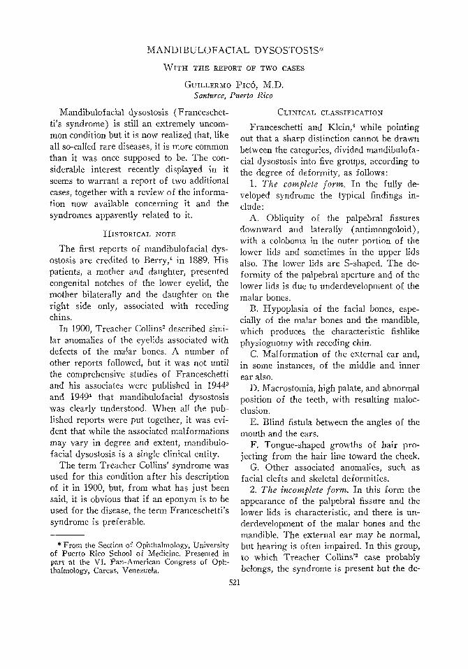
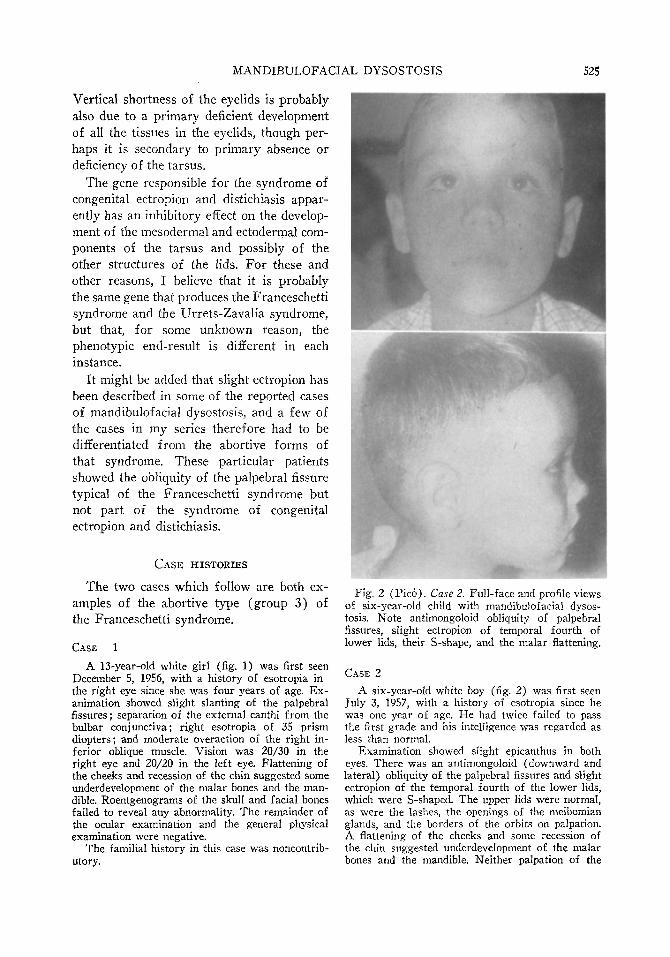
Fig. 1 (Pico). Case 1. Full-face and profile views of 13-year-old girl with mandibulofacial dysostosis. Note slight slanting of palpebral fissures and apparent underdevelopment of malar bones and mandible shown by flattening of cheeks and recession of chin.
distichiasis. Two persons had distichiasis alone. Previously reported cases of ectropion, which numbered five at most, lacked many of the characteristics of the anomaly observed in this family. Similarly, reported cases of associated ectropion and distichiasis, which number two at most, lacked other characteristic features of the cases that I reported.
Histologic examination of specimens obtained in two cases in this family showed
absence of meibomian glands and either complete absence of the tarsal plate or the presence of only vestigial structures. Absence, weakness, and other defects of the tarsus were determined by palpation in the other cases in my reported genealogy, but a clinical examination, of course, cannot be regarded as conclusive. This special etiologic factor was not investigated in any of the cases of ectropion previously reported.
Other characteristic features of the syndrome of congenital ectropion and distichiasis included narrowing of the palpebral fissure horizontally, which was described by the patients as "small eyes," and vertical shortening of the eyelids.
The mode of inheritance in this congenital syndrome is evidently of the autosomal dominant type. The responsible gene in the series showed a high degree of penetrance; more than half of the members of the family presented one defect or the other or both. The genetic expression, however, was somewhat variable; the degree of ectropion or distichiasis varied widely. On the other hand, there was no doubt of the expression, since, of the 10 living affected members of the family who could be examined, only two showed distichiasis alone. The other eight showed both ectropion and distichiasis.
In the 10 living patients in my series who presented ectropion, the anomaly involved all four lids in four cases and was complete in the lower lids, but less marked in the upper lids, in every case. One patient had complete ectropion of both lower lids, with normal upper lids. The other five patients had only slight ectropion of both lower lids. Ectropion of the upper eyelids is not a feature of mandibulofacial dysostosis.
No bony abnormalities were seen grossly or elicited by palpation in any of these cases. The borders of the orbit were normal, and no obliquity of the palpebral fissures was noted. In this syndrome, the ectropion and distichiasis are evidently the result of a defect of tarsal development and have nothing to do with anomalies of the facial bones.
MANDIBULOFACIAL DYSOSTOSIS 525
Vertical shortness of the eyelids is probably also due to a primary deficient development of all the tissues in the eyelids, though perhaps it is secondary to primary absence or deficiency of the tarsus.
The gene responsible for the syndrome of congenital ectropion and distichiasis apparently has an inhibitory effect on the development of the mesodermal and ectodermal components of the tarsus and possibly of the other structures of the lids. For these and other reasons, I believe that it is probably the same gene that produces the Franceschetti syndrome and the Urrets-Zavalia syndrome, but that, for some unknown reason, the phenotypic end-result is different in each instance.
It might be added that slight ectropion has been described in some of the reported cases of mandibulofacial dysostosis, and a few of the cases in my series therefore had to be differentiated from the abortive forms of that syndrome. These particular patients showed the obliquity of the palpebral fissure typical of the Franceschetti syndrome but not part of the syndrome of congenital ectropion and distichiasis.
CASE HISTORIES
The two cases which follow are both examples of the abortive type (group 3) of the Franceschetti syndrome.
CASE 1
A 13-year-old white girl (fig. 1) was first seen December 5, 1956, with a history of esotropia in the right eye since she was four years of age. Ex-animation showed slight slanting of the palpebral fissures; separation of the external canthi from the bulbar conjunctiva; right esotropia of 35 prism diopters; and moderate overaction of the right inferior oblique muscle. Vision was 20/30 in the right eye and 20/20 in the left eye. Flattening of the cheeks and recession of the chin suggested some underdevelopment of the malar bones and the mandible. Roentgenograms of the skull and facial bones failed to reveal any abnormality. The remainder of the ocular examination and the general physical examination were negative.
The familial history in this case was noncontrib-utory.
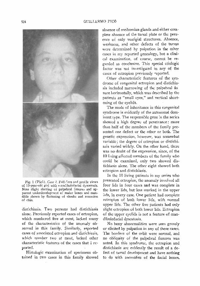
Fig. 2 (Pico) . Case 2. Full-face and profile views of six-year-old child with mandibulofacial dysostosis. Note antimongoloid obliquity of palpebral fissures, slight ectropion of temporal fourth of lower lids, their S-shape, and the malar flattening.
CASE 2
A six-year-old white boy (fig. 2) was first seen July 3, 1957, with a history of esotropia since he was one year of age. He had twice failed to pass the first grade and his intelligence was regarded as less than normal.
Examination showed slight epicanthus in both eyes. There was an antimongoloid (downward and lateral) obliquity of the palpebral fissures and slight ectropion of the temporal fourth of the lower lids, which were S-shaped. The upper lids were normal, as were the lashes, the openings of the meibomian glands, and the borders of the orbits on palpation. A flattening of the cheeks and some recession of the chin suggested underdevelopment of the malar bones and the mandible. Neither palpation of the
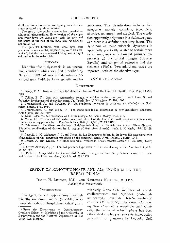
526 GUILLERMO PICO
skull and facial bones nor roentgenograms of these areas revealed any abnormalities.
The rest of the ocular examination revealed no additional abnormalities. Examination of the upper and lower jaws, the palate and teeth, the ears, and the bones of the rest of the body also revealed no abnormalities.
The patient's brothers, who were aged four years and seven months, respectively, were also examined, but the only abnormal finding was a slight epicanthus in the older child.
S U M M A R Y
Mandibulofacial dysostosis is an uncommon condition which was first described by Berry in 1889 but was not definitively described until 1949, by Franceschetti and his
INTRODUCTION
The agent, 2-diethoxyphosphinylthioethyl-trimethylammonium iodide (217-MI ; echo-thiophate iodide; phospholine iodide), is a
* From the Department of Ophthalmology, Graduate School of Medicine of the University of Pennsylvania and the Research Department of The Wills Eye Hospital,
associates. The classification includes five categories, namely, complete, incomplete, abortive, unilateral, and atypical. The condition apparently originates in a defective gene, and there is a definite hereditary factor. The syndrome of mandibulofacial dysostosis is apparently genetically related to certain other syndromes, especially familial primary hy-poplasia of the orbital margin (Urre t s -Zavalia) and congenital ectropion and dis-tichiasis ( P i c o ) . Two additional cases are reported, both of the abortive type.
1475 Wilson Avenue.
relatively irreversible inhibitor of acetyl-cholinesterase1 and N,N'-bis (2-diethyl-aminoethyl) oxamide bis-2-chlorobenzyl chloride ( W I N 8077; ambenonium chloride; mytelase chloride) a reversible one.2 Clinically the value of echothiophate has been established amply, ever since its introduction in control of glaucoma by Leopold, Gold
REFERENCES
1. Berry, F. A.: Note on a congenital defect (coloboma?) of the lower lid. Ophth. Hosp. Rep., 12:255, 1889.
2. Collins, E. T.: Case with symmetrical congenital notches in the outer part of each lower lid and defective development of the malar bones. Tr. Ophth. Soc. U. Kingdom, 20:190, 1900.
3. Franceschetti, A., and Zwahlen, P.: Un syndrome nouveau: la dysostose mandibulofaciale. Bull. suisse d. med, 1:60, 1944.
4. Franceschetti, A., and Klein, D.: The mandibulo-facial dysostosis: A new hereditary svndrome. Acta Ophth, 27:143, 1949.
5. Duke-Elder, W. S.: Textbook of Ophthalmology. St. Louis, Mosby, 1952, v. 5. 6. Mann, I.: Deficiency of the malar bones with defect of the lower lid; with notes of a similar case,
treatment and suggestions by T. Pomfret Kilner. Brit. J. Ophth, 27:13, 1943. 7. Debusmann: Familiare kombinierte Gesichtsmissbildung in Bereich des ersten Viszeralbogens.
(Familial combination of deformities in region of first visceral arch). Arch. f. Kinderh, 120:133-139, 1940.
8. Leopold, I. H , Mahoney, J. F , and Price, M. L.: Symmetric defects in the lower lids associated with abnormalities of the zygomatic processes of the temporal bones. Arch. Ophth, 34:210, 1945.
9. Brohm, F , and Kluska, V.: Mandibulofacial dysostosis (Franceschetti-Zwahlen) Lek. listy, 2:329, 1947.
10. Urrets-Zavalia, A , Jr.: Familial primary hypoplasia of the orbital margin. Tr. Am. Acad. Ophth, 59:42, 1955.
11. Pico, G.: Congenital ectropion and distichiasis: Etiologic and hereditary factors: a report of cases and review of the literature. Am. J. Ophth, 47:363, 1959.
E F F E C T O F E C H O T H I O P H A T E A N D A M B E N O N I U M O N T H E R A B B I T P U P I L *
IRVING H. LEOPOLD, M.D., AND NARENDRA K R I S H N A , M.B.B.S. Philadelphia, Pennsylvania