54595681-embriologi
TRANSCRIPT

EMBRIOLOGI UMUM
Dr. Thontowi Djauhari NS, MKes
Laboratorium Anatomi
Program Pendidikan Dokter
Universitas Muhammadiyah Malang

Awalnya manusia mempunyai 46 kromos (diploid)
Proses Meiosis akan mengurangi jumlah sel menjadi 23 kromosom (haploid)
Penyatuan ovum + sperma akan menghasilkan 46 kromosom
MEIOSIS FERTILIZATION
Haploid gametes (n = 23)
Egg cell haploid
Sperm cell haploid
Diploidzygote
(2n = 46)Multicellular
diploid adults (2n = 46)
Mitosis anddevelopment

GAMETOGENESISGAMETOGENESIS
PEMBENTUKAN PEMBENTUKAN SEL SEKS PRIA SEL SEKS PRIA DAN WANITA DAN WANITA ATAU SEL GAMET ATAU SEL GAMET YANG BERASAL YANG BERASAL DARI GERM CELLSDARI GERM CELLS
OOGENESIS OOGENESIS TERJADI PADA TERJADI PADA WANITAWANITA
SPERMATOGENESISPERMATOGENESIS S TERJADI PADA TERJADI PADA PRIAPRIA

Testis and Formation of Sperm
2n2n
nn
2n2n
nn
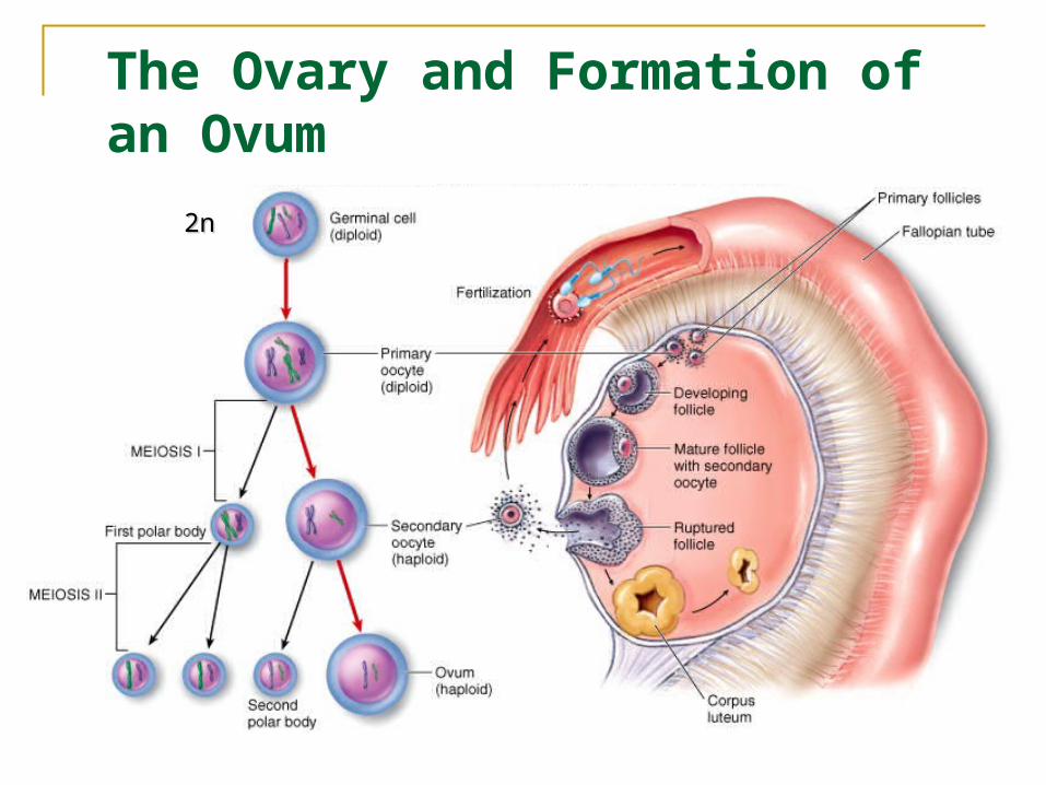
The Ovary and Formation of an Ovum
2n2n


GAMETOGENESIS
MITOSIS: MENJADI 2 SEL YANG SAMA
MEIOSIS : I : - PAIRING KROMOSOM HOMOLOG
- CROSS OVER (PERTUKARAN SEGMEN)
II: - SINTESIS DNA TIDAK TERJADI
- PEMISAHAN KROMOSOM GANDA MENJADI TUNGGAL

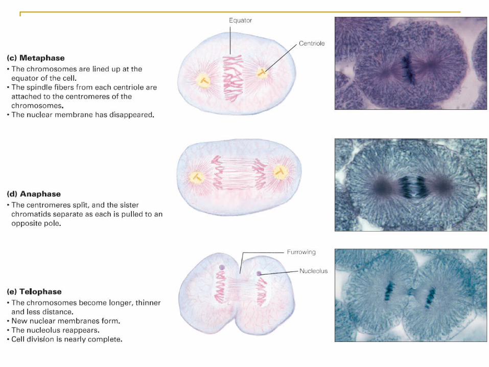
MITOSIS
Mitosis is a continuum but biologists distinguish 4 stages
Prophase Metaphase Anaphase Telophase


MEIOSIS
Percampuran materi genetis pada waktu cross over sehingga dapat terjadi variasi genetis
Supaya sel kelamin menjadi kromosom haploid dengan jumlah DNA ½ dari jumlah DNA sel somatis (meiosis 2)
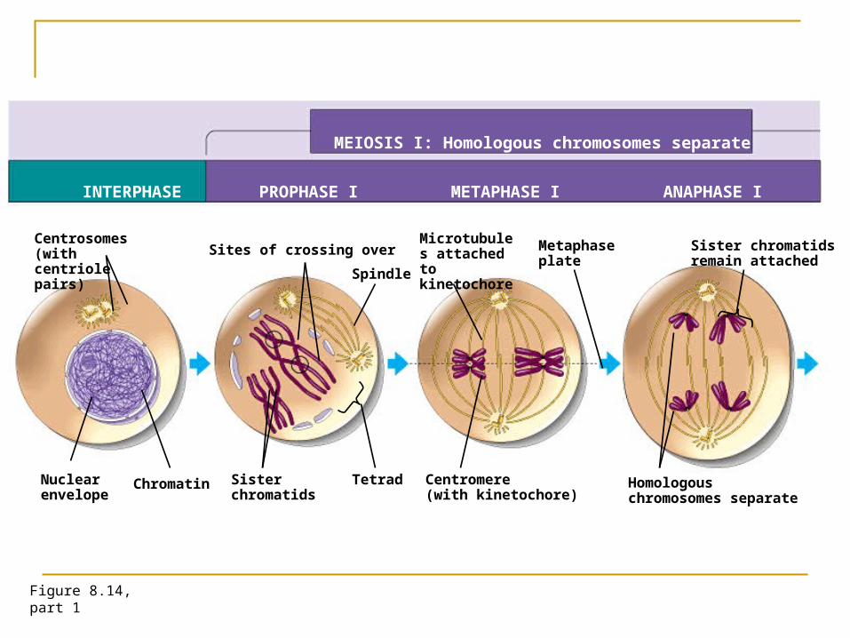
Figure 8.14, part 1
MEIOSIS I: Homologous chromosomes separate
INTERPHASE PROPHASE I METAPHASE I ANAPHASE I
Centrosomes(withcentriolepairs)
Nuclearenvelope
Chromatin
Sites of crossing over
Spindle
Sisterchromatids
Tetrad
Microtubules attached tokinetochore
Metaphaseplate
Centromere(with kinetochore)
Sister chromatidsremain attached
Homologouschromosomes separate
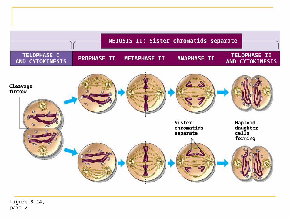
Figure 8.14, part 2
MEIOSIS II: Sister chromatids separate
TELOPHASE IAND CYTOKINESIS PROPHASE II METAPHASE II ANAPHASE II
Cleavagefurrow
Sister chromatidsseparate
TELOPHASE IIAND CYTOKINESIS
Haploiddaughter cellsforming

KELAINAN NON DYSJUNCTION
Non dysjunction dapat terjadi pada waktu meiosis 1 atau meiosis 2
Turner Syndrome 45,XO(female)
Trisomy X 47, XXX (female)
Klinefelter Syndrome 47,XXY (male) Extra “Y” chromosome 47,XYY (male)

Nondisjuction

Down syndrome:trisomy for Chr 21 (47 Mb)
normal disjoining
non-disjoining
non-disjoining
•trisomy of chromosome number 21 (1 in 700 births)—mental retardation, mongoloid features, and heart defects

XO – Turner Syndrome
Turner Syndrome (XO), Incidence: 1 in 2500 female births•Females missing one X chromosome (XO)

XXY – Klinefelter Syndrome
Klinefelter Syndrome (XXY), Incidence: 1:1000 male births•Males with an extra X chromosome (XXY) (1 in 1000 male births)
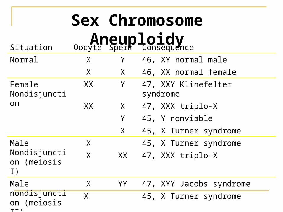
Sex Chromosome Aneuploidy
Situation Oocyte Sperm Consequence
Normal X Y 46, XY normal male
X X 46, XX normal female
Female Nondisjunction
XX Y 47, XXY Klinefelter syndrome
XX X 47, XXX triplo-X
Y 45, Y nonviable
X 45, X Turner syndrome
Male Nondisjunction (meiosis I)
X 45, X Turner syndrome
X XX 47, XXX triplo-X
Male nondisjunction (meiosis II)
X YY 47, XYY Jacobs syndrome
X 45, X Turner syndrome

FERTILISASIDEFINISI : Fertilisasi adalah proses fusi antara
nukleus spermatosoa dengan ovum
Fertilisasi dimulai dengan reaksi akrosom dari Spermatosoa dan diakhiri dengan aktivasi Oocyt
Selama di dalam tractus genitalis perempuan,sebelum fertilisasi, spermatosoa mengalami : Kapasitasi: pelepasan glikoprotein dan protein plasma Reaksi akrosom: pelepasan ensim untuk mencairkan
corona radiata dan zona pellucida

A : Belum Kapasitasi
B : Kapasitasi
C : Reaksi Akrosom
C
BA


FERTILISASI
Bila 1 spermatosoa masuk ke dalam nukleus
ovum, maka terjadi reaksi ovum, membrana oosit tidak dapat ditembus sperma lagi
Nukleus sperma (23 kromosom) fusi dengan nukleus oosit (23 kromosom) menjadi sigot (46 kromosom)
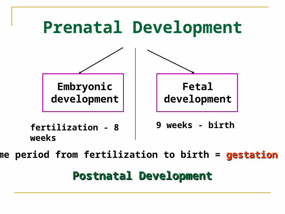
Prenatal Development
Embryonic development
fertilization - 8 weeks
Fetal development
9 weeks - birth
Postnatal DevelopmentPostnatal Development
time period from fertilization to birth = gestationgestation

Development Overview
frog egg
frog sperm
midsectional views
top view side view
Organs increase in size and gradually assume specialized functions.
Eggs form and mature in female reproductive organs, and sperm form and mature in male reproductive organs.
A sperm and an egg fuse at their plasma membrane, then the nucleus of one fuses with the nucleus of the other to form the zygote.
By a series of mitotic cell divisions, different daughter cells receive different regions of the egg cytoplasm.
Cell divisions, migrations, and rearrangements produce two or three primary tissues, the forerunners of specialized tissues and organs.
Subpopulations of cells are sculpted into specialized organs and tissues in prescribed spatial patterns at prescribed times.
Gamete formation
Fertilization
Cleavage
Gastrulation
Organ formation
Growth, tissuespecialization
Neurulation
Morula BlastocystImplantation

PERKEMBANGAN EMBRIO MINGGU PERTAMA
Periode ovulasi sampai implantasi Berlangsung ± 6 hari Sigot mengalami pembelahan sel:
2 sel 4 sel 8 sel 16 sel (morula) Saat nampak lubang (vacuola) pada
perkembangan morula : free blastocyst

Blastocystwith blastocoele cavity
Morula solid ball of cells
CleavageEarly division of zygote into multiple cells without increase in size, partitions contents
Zygote

Fig 28-3
Periode implantasiPeriode implantasi
Berlangsung mulai hari ke 6 saat Berlangsung mulai hari ke 6 saat melekatnya blastocyst pada epithel melekatnya blastocyst pada epithel endometrium sampai hari ke 12 endometrium sampai hari ke 12 setelah ovulasisetelah ovulasi
Terdapat 2 kelompok sel Terdapat 2 kelompok sel inner cells inner cells massmass yang disebut: yang disebut: ectoderm dan ectoderm dan endodermendoderm
Pada bagian luar ECTODERM Pada bagian luar ECTODERM terdapat kelompok sel yang terdapat kelompok sel yang dinamakan dinamakan trophoblasttrophoblast
Terdiri dari :Terdiri dari :CytotrophoblastCytotrophoblastSyncytiotrophoblastSyncytiotrophoblast
Pada periode ini mulai terbentuk Pada periode ini mulai terbentuk amniotic cavityamniotic cavity

Periode : Gastrulation
Day 10-11: cells move inward
Forms 3 layers:
• Ectoderm• Mesoderm• Endoderm
Will become:
Epidermis, CNS, sense organs, neural crest
Skeleton, muscles,Blood vessels, heart, gonads
Lining of GI & air tracts, liver, pancreas
►Pembentukan primitive streak pada permukaan ectodermPembentukan primitive streak pada permukaan ectoderm
►Antara ectoderm dan endoderm terdapat sel mesenchyme Antara ectoderm dan endoderm terdapat sel mesenchyme yang berdiferensiasi menjadi mesoderm (intraembryonic yang berdiferensiasi menjadi mesoderm (intraembryonic mesoderm)mesoderm)
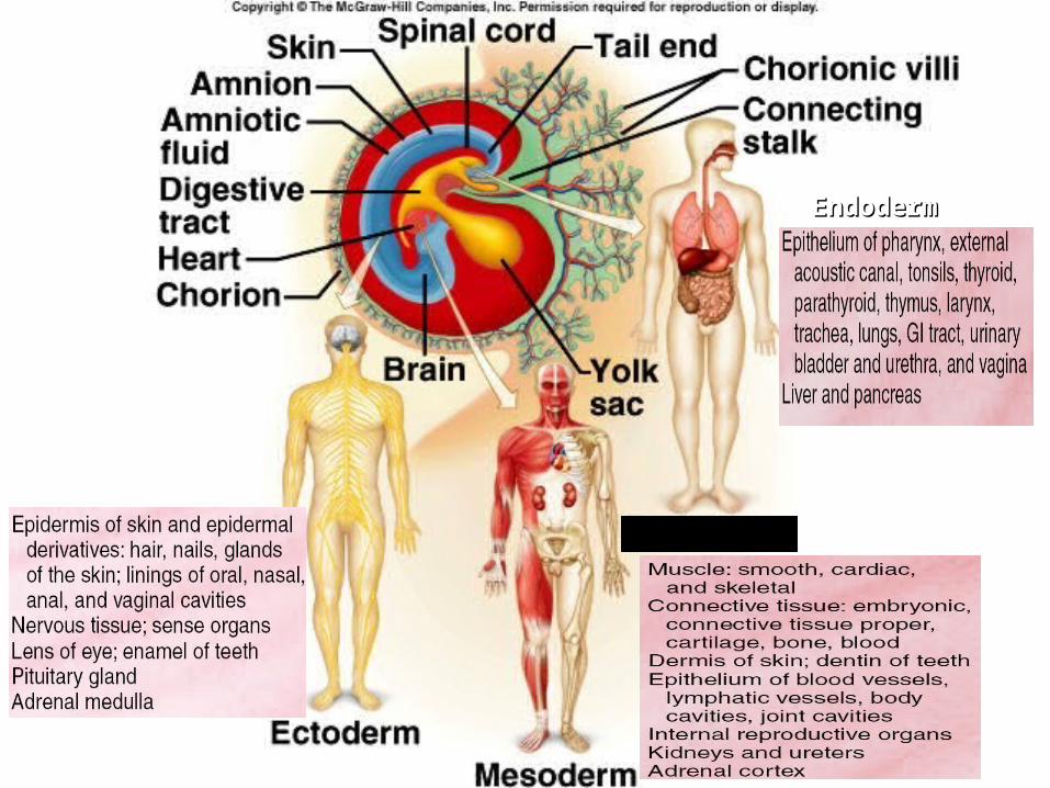
EndodermEndoderm

Human Chorionic Gonadotropin (HCG)
Pengeluaran hormon saat blastocytst by the blastocyst
Stimulates corpus luteum to keep making progesterone and estrogens
This maintains endometrium, prevents menstruation
Can be detected by week 3 with a home pregnancy test

KELAINAN
Abortus spontan Implantasi yang
abnormal Mola hydatidosa/
choriocarcinoma

PERKEMBANGAN EMBRIO MINGGU KETIGA
Intra embryonic mesoderm meluas, bersatu dengan extraembryonic mesoderm
Pembentukan villi dari trophoblast Akhir minggu ke 3 mesoderm berdiferensiasi
menjadi pembuluh darah villous capillary system
Pembentukan neural plate neural tube Pembentukan neural crest dari ectoderm
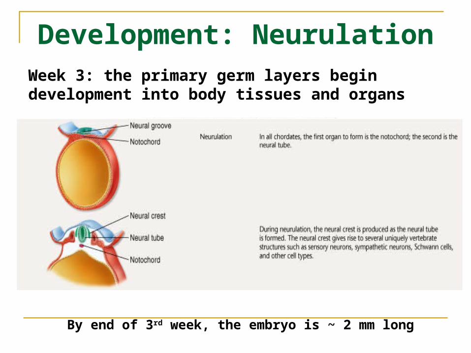
Development: NeurulationWeek 3: the primary germ layers begin development into body tissues and organs
By end of 3rd week, the embryo is ~ 2 mm long

Neurulation Development of hollow nerve cord Neural groove forms
paired neural folds
somites
pharyngealarches

KELAINANTeratoma sacrococcygeal (sisa primitive streakNeural tube defect (meningocele dll)

PERIODE FETAL►4th week = 4th week = organogenesisorganogenesis
►Embryo ~ 5 mmEmbryo ~ 5 mm
►Critical time in developmentCritical time in development
Second Month Embryo ~ 25 mm
7 weeks
Great changes occur in morphology Limbs assume adult shape Major internal organs
evident
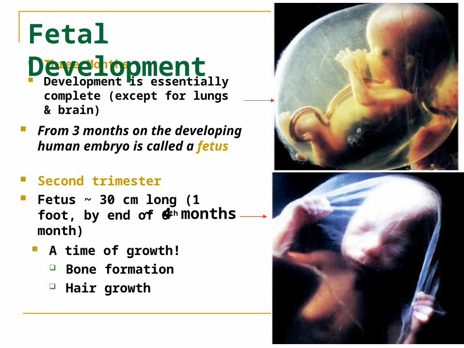
Three Months Development is essentially
complete (except for lungs & brain)
Fetal Development
From 3 months on the developing human embryo is called a fetus
Second trimester Fetus ~ 30 cm long (1 foot, by
end of 6th month)
A time of growth! Bone formation Hair growth
~ 4 months

Placenta
Third trimester Weight ~ doubles
Major change is great increase in size Most major nerve
tracts formed in brain Nutrients from
mother’s blood via placenta
Fetal Development

Placental-Fetal circulation

Sesungguhnya kami (Allah) menciptakan manusia dalam bentuk yang sebaik-baiknya
QS 95:4

TWIN


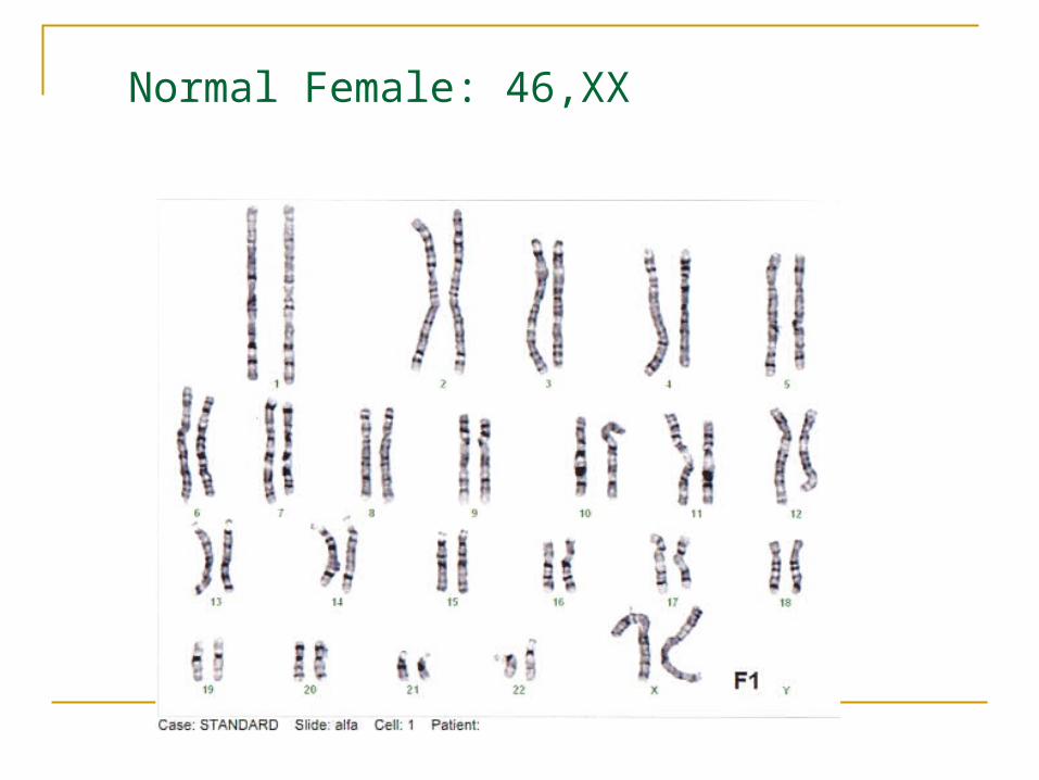
Normal Female: 46,XX

Normal Male: 46,XY

Autosomal Abnormalities

Trisomy 21 Down Syndrome 47,XY,+21
47, XX, 21+ 47, XX, 21+ Female with Down SyndromeFemale with Down Syndrome
47, XY, 21+47, XY, 21+ Male with Down SyndromeMale with Down Syndrome

Trisomy 21 Major Clinical Features
mental retardation slanted palpebral
fissures epicanthal folds small, round, flat face small mouth, protruding
tongue congenital heart
problems
Brushfield spots (iris)
small, hypoplastic ears
simian creases hypotonia, lax joints,
hyperextensive

Trisomy 13 Patau Syndrome 47,XY,13+

Trisomy 13 Major Clinical Features
mental retardation growth retardation microcephaly cleft lip/palate small jaw (micrognathia) deformed, low-set ears
polydactyly congenital heart
defects rocker bottom feet seizures low birth weight
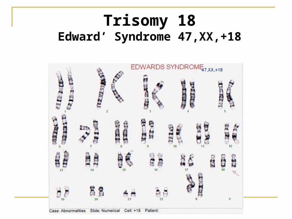
Trisomy 18 Edward’ Syndrome 47,XX,+18

Trisomy 18 Major Clinical Features
mental retardation growth retardation short neck cleft lip/palate dislocated
hips/abnormal pelvis deformed, low-set
ears
hypertonia congenital heart
disease horseshoe kidneys hydronephrosis short sternum pyloric stenosis

Cri du chat Syndrome (5p-)

Cri du chat Major Clinical Features
distinctive cat-like cry
profound developmental retardation
severe mental retardation
microcephaly hypotonia
hypertelorism congenital heart
disease round, moon-shaped
face large mouth, short
philtrum low set ears hand and foot
abnormalities

Sex Chromosome Anomalies
General features:Some growth retardation (GR)
Reproductive anomalies/problemsGood viabilityPrenatally diagnosableAssociated with spontaneous abortion (Sab)
HBD/CA/Sex
Sex Chromosome Anomalies
Monosomy X: Turner’s Syndrome (45, X) Trisomy X: Triplo-X Syndrome (47, XXX) Trisomy (47, XXY): Klinefelter’s Syndrome Trisomy (47, XYY): XYY Syndrome

Turner’s Syndrome 45,X
Turner’s Syndrome Major Clinical Features
female phenotype short (less than 5 feet) primary amenorrhea low estrogen levels maldevelopment of the
ovaries sterility
webbing of the skin of the neck
wide-spaced nipples
edema at birth cardiovascular
problems
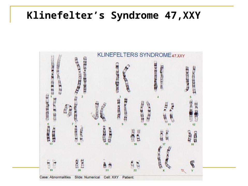
Klinefelter’s Syndrome 47,XXY
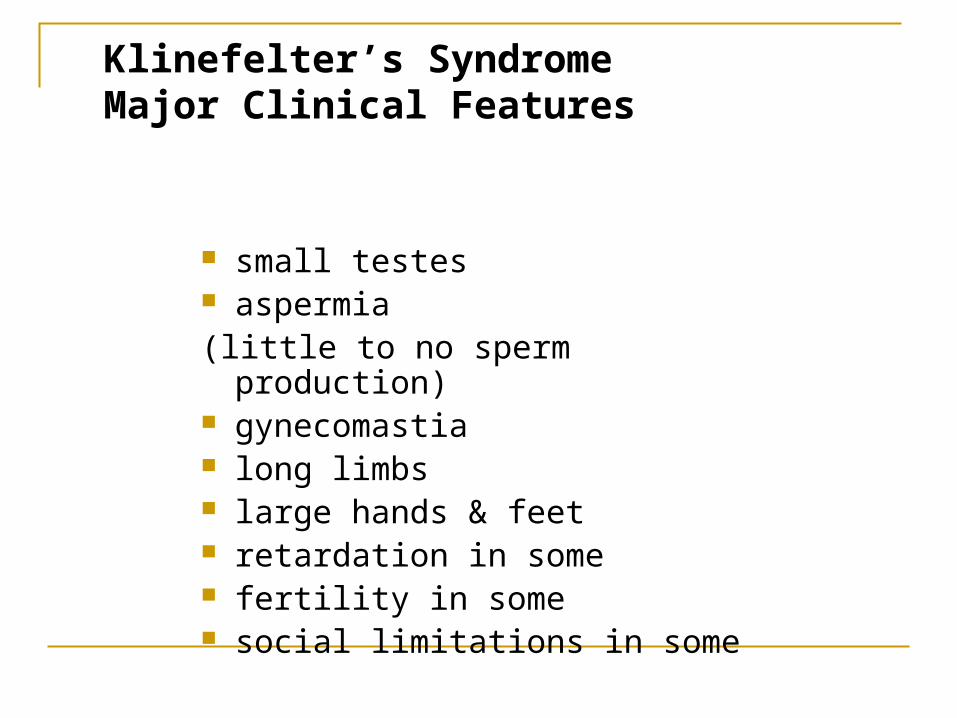
Klinefelter’s Syndrome Major Clinical Features
small testes aspermia (little to no sperm production) gynecomastia long limbs large hands & feet retardation in some fertility in some social limitations in some