growth development system
DESCRIPTION
Growth development systemTRANSCRIPT
Infeksi virus
R. Lia Kusumawati, dr, MS, SpMK
Departemen Mikrobiologi
FK USU

1. Virus Herpes Struktur & morfologi• Virion: spheris, icosahedral capsid• Genom : linear, DNA untai ganda• Protein: > 35 jenis protein pada virion• Envelope: glikoprotein, reseptor Fc• Replikasi: nucleus bertunas dari membran nuclear.
• Ciri khas infeksi virus herpes:
1.Menyebabkan infeksi laten
2. Menetap secara indefenitif pada penderita
3. Sering reaktif pada immunosuppressed host

Infeksi virus herpes
1. Virus herpes simpleks tipe 1
- hanya pada manusia, tidak memiliki vektor/reservoir hewan
- Infeksi laten: Sacral ganglia
- Usia terinfeksi: anak balita
- Transmisi/ penularan: kontak langsung, saliva
- Gejala Klinis:
a. Inf. Primer:gingingostomatitis, pharyngotonsilitis, keratoconjuctivitis, inf neonatal.
b. Recurent inf: fever, keratitis.
c. Inf primer/ recurrent: Herpes kulit di atas pinggang, pd tangan, siku, herpatic withlow, eczema herpeticum, genital herpes, herpes encephalitis, herpes meningitis.
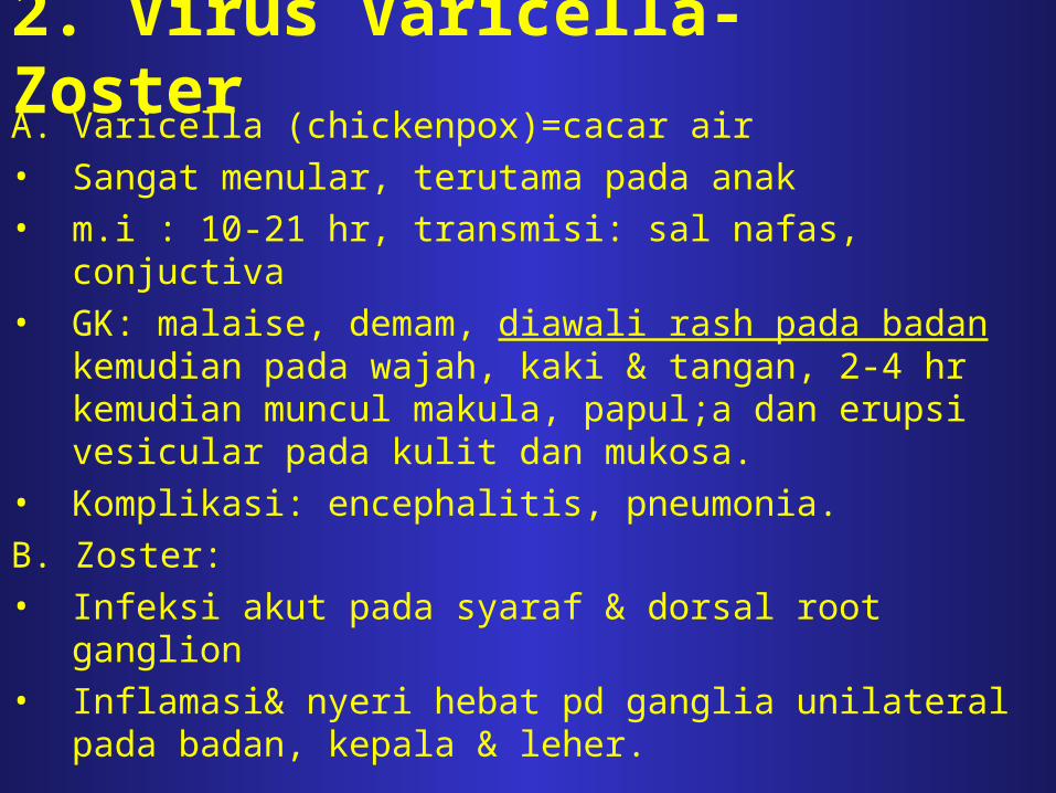
2. Virus Varicella-ZosterA. Varicella (chickenpox)=cacar air• Sangat menular, terutama pada anak• m.i : 10-21 hr, transmisi: sal nafas, conjuctiva• GK: malaise, demam, diawali rash pada badan kemudian
pada wajah, kaki & tangan, 2-4 hr kemudian muncul makula, papul;a dan erupsi vesicular pada kulit dan mukosa.
• Komplikasi: encephalitis, pneumonia.
B. Zoster:• Infeksi akut pada syaraf & dorsal root ganglion• Inflamasi& nyeri hebat pd ganglia unilateral pada badan,
kepala & leher.

Terapi Herpes Virus• HSV 1 dan Varicella-Zoster virus:
-Acyclovir
-Vidarabin

2. POXVIRIDAE
Structure & composition:- Oval or brick shaped, 400nm (length) x 230nm ()
external surface shows ridges, contains core & lateral
bodies
- Composition: DNA (3%),protein (90%), lipid(5%)
- Genome: linear double stranded DNA, MW 85-150x106
rich in adenine & thymine bases, low
guanine-plus-cytosine
- Contain >100 polypeptides → many enzimes in core
- Envelope (+)

VACCINIA & VARIOLA(Poxvirus infection in humans)
• 1798 : Jenner introduced vaccination with live
virus
• 1967 : WHO worldwide campaign to
eradicate smallpox
• 1980 : smallpox was officially declared
eliminated

Composition of vaccinia & variola viruses:
# Host :
- variola : only humans & monkeys
- vaccinia : broad host range, including
rabbits & mice
# Grow on chorioallantoic membrane of 10-12 days
old chick embryo, but variola produces much
smaller pocks
# Grow in several types of chick & primate cell
lines

Pathogenesis of smallpox:
- Portal of entry : mucous membranes of upper
respiratory tract
- After viral entry :
1. primary multiplication in the lymphoid tissue
draining the site of entry
2. transient viremia and infection of reticulo-
endothelial cells throughout the body
3. secondary phase of multiplication in those cells
4. secondary, more intense viremia
5. clinical disease

Clinical findings :
Incubation period of variola (smallpox) = 12 days
1. Fever & malaise : 1-5 days
2. Exanthems appear : - papular (1-4 days)
- vesicular (1-4 days)
- pustular (2-6 days)
3. Crusts formed (2-4 weeks after 1st sign of lesion)
and leaving pink that faded slowly

Laboratory diagnosis:
a. Isolation & identification :
- skin lesions specimen (choice for viral isolation)
- direct exam (electron microscope) :
rapid identification of viral particles (± 1 hr)
differentiate poxvirus infection from
chickenpox
- viral isolation is carried out by inoculation of
vesicular fluid onto the chorioallantoic
membrane of chick embryos
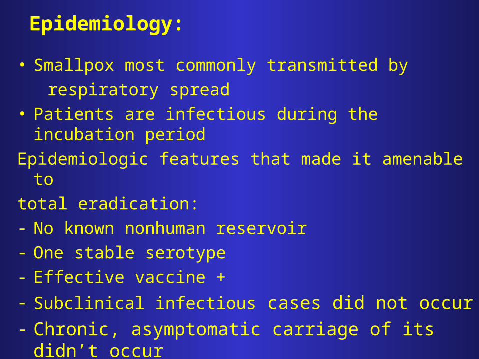
Epidemiology:
• Smallpox most commonly transmitted by
respiratory spread• Patients are infectious during the incubation period
Epidemiologic features that made it amenable to
total eradication:- No known nonhuman reservoir- One stable serotype- Effective vaccine +
- Subclinical infectious cases did not occur
- Chronic, asymptomatic carriage of its didn’t occur

3. ENTEROVIRUSES
• Enteroviruses are a genus of the Picornavirus family which replicate mainly in the gut.
• Single stranded naked RNA virus with icosahedral symmetry
• Unlike rhinoviruses, they are stable in acid pH, enables to survive exposure to gastric acid.
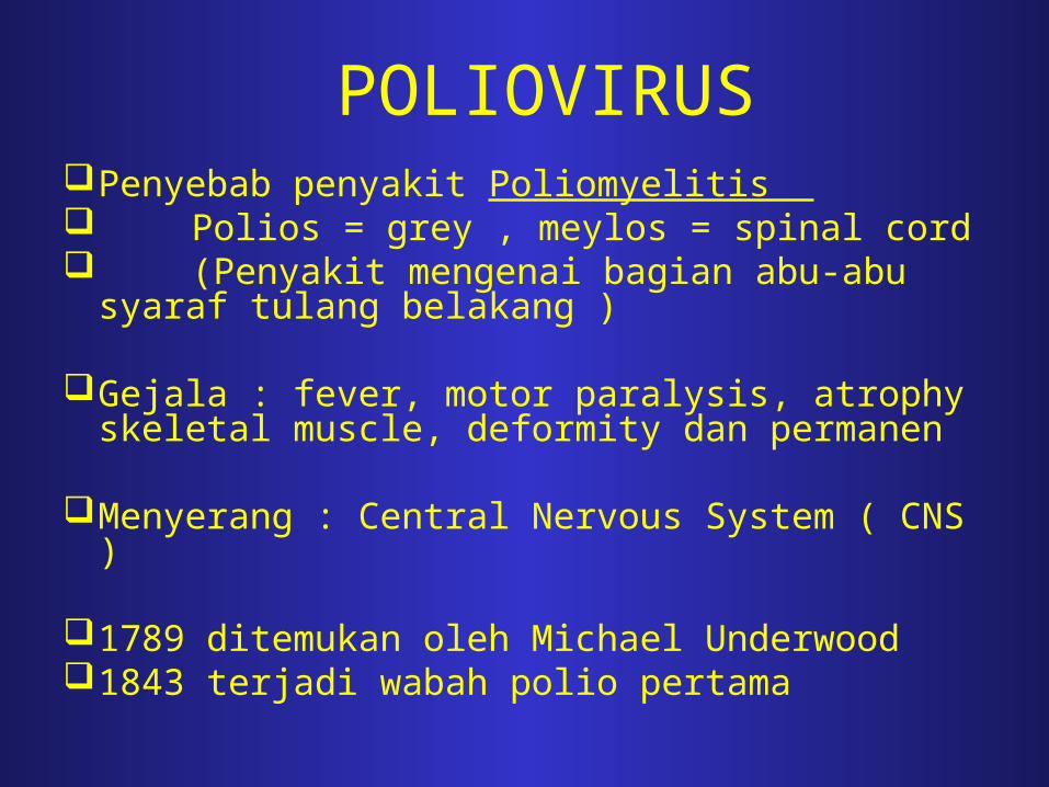
POLIOVIRUSPenyebab penyakit Poliomyelitis Polios = grey , meylos = spinal cord (Penyakit mengenai bagian abu-abu syaraf
tulang belakang )
Gejala : fever, motor paralysis, atrophy skeletal muscle, deformity dan permanen
Menyerang : Central Nervous System ( CNS )
1789 ditemukan oleh Michael Underwood1843 terjadi wabah polio pertama
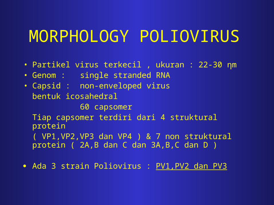
MORPHOLOGY POLIOVIRUS
• Partikel virus terkecil , ukuran : 22-30 ηm• Genom : single stranded RNA • Capsid : non-enveloped virus
bentuk icosahedral 60 capsomer
Tiap capsomer terdiri dari 4 struktural protein ( VP1,VP2,VP3 dan VP4 ) & 7 non struktural protein ( 2A,B dan C dan 3A,B,C dan D )
Ada 3 strain Poliovirus : PV1,PV2 dan PV3

SIFAT POLIOVIRUS
I. Resisten terhadap :
1. pH rendah ( pH=3 ), asam lambung ( gastric acid ), bile virus dapat penetrasi sampai ke intestinum menginfeksi multiplikasi di epithelium & mesenteric lymph node
2. Beberapa enzym proteolytic
3. Beberapa desinfectan : alcohol 70%, lisol 25%, eter, desoxycholate
4. Berbagai jenis detergen

II. Inaktif terhadap :
1. Formaldehyde (0,3%), HCl 0,1 N dan halogen lainnya
2. Pengeringan, cahaya, panas ( 500 selama 1 jam )
3. Larutan Glutaraldehyde : sangat cepat menginaktifkan poliovirus
4. Virus mati pada Pasteurisasi ( 620-720C )
5. Virus dalam spinal cord mati pd pH 12 atau pH > 11
SIFAT POLIOVIRUS

MODE OF TRANSMISSIONPoliovirus transmisi ke orang lain melalui :
1. Direct contact : fecal-oral contact 2. Indirect contact : melalui makanan dan air yang terkontaminasi oleh faeces atau saliva
Pasien dalam keadaan infectious :• Masa inkubasi : 1-2 minggu - 7-10 hari sebelum & sesudah adanya tanda penyakit - Virus dalam tenggorokan ( throat ) ± 1 minggu sesudah timbul
gejala diekskresikan dlm faeces - Virus dapat berada dalam feces selama beberapa minggu
sampai beberapa bulan - Infeksi yang diperoleh dapat memberikan Life long immunity
( kekebalan sepanjang hidup )
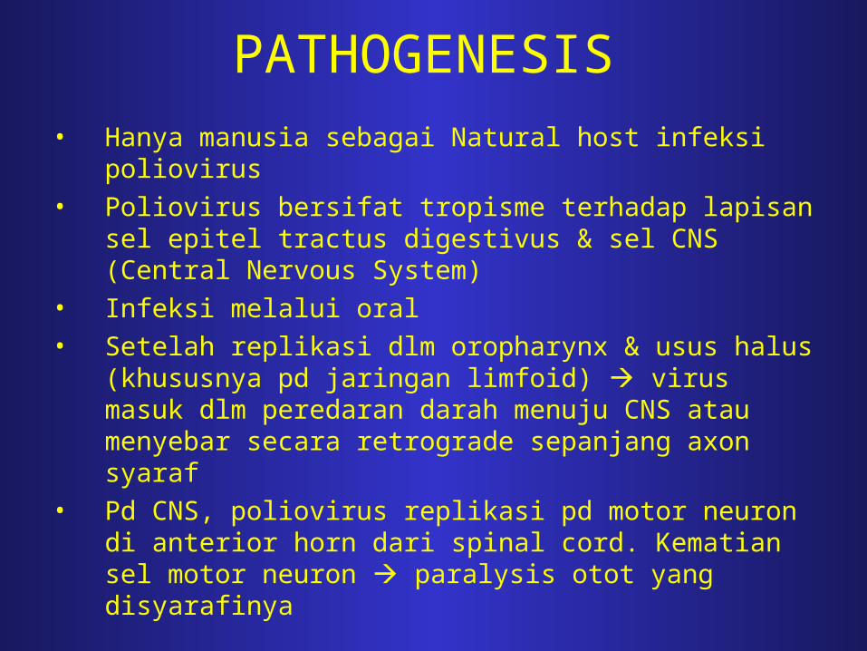
PATHOGENESIS
• Hanya manusia sebagai Natural host infeksi poliovirus
• Poliovirus bersifat tropisme terhadap lapisan sel epitel tractus digestivus & sel CNS (Central Nervous System)
• Infeksi melalui oral
• Setelah replikasi dlm oropharynx & usus halus (khususnya pd jaringan limfoid) virus masuk dlm peredaran darah menuju CNS atau menyebar secara retrograde sepanjang axon syaraf
• Pd CNS, poliovirus replikasi pd motor neuron di anterior horn dari spinal cord. Kematian sel motor neuron paralysis otot yang disyarafinya

PATOGENESIS INFEKSI POLIOVIRUS
Replikasi Replikasi Replikasi
Alimentary phase Lymphatic phase Viremic phase Neurologic phase
Mucosa Tonsil Blood - Anterior hornoropharynx Lymph node Circulation spinal cord
(deep cervical) - Dorsal rootganglia - Motor neuron
VIRUS - Brain stem - Terjadi CPE
Mucosa Peyer patches Blood - Internal organ Intestinal Lymph node Circulation - Lymph node regional
mesenterium
Peripheral - Brain nerve - Skeletal muscle
- Terjadi CPE

POLIOVIRUS
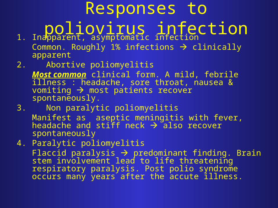
Responses to poliovirus infection1. Inapparent, asymptomatic infection
Common. Roughly 1% infections clinically apparent2. Abortive poliomyelitis
Most common clinical form. A mild, febrile illness : headache, sore throat, nausea & vomiting most patients recover spontaneously.
3. Non paralytic poliomyelitisManifest as aseptic meningitis with fever, headache and stiff neck also recover spontaneously
4. Paralytic poliomyelitisFlaccid paralysis predominant finding. Brain stem involvement lead to life threatening respiratory paralysis. Post polio syndrome occurs many years after the accute illness.

REPLICATION & POLIOVIRUS LIFE CYCLE
I. Poliovirus interaction with poliovirus receptor (PVR)
Attachment
II. Uncoating
III. Protein Synthesis
IV. Protein Processing
V. RNA Replication
VI. Packaging and Release


DIAGNOSIS LABORATORIUM
1.Isolasi virus pilihan utama diagnosis2. Pemeriksaan titer Antibody utk individu immunocompromised
- Spesimen untuk isolasi poliovirus berasal dari :1.Throat swab2. Throat washing3. Cerebrospinal fluid jarang diperoleh4. Darah ( waktu viremia phase : 6-9 hari sesudah infeksi )5. Feces
- Biakan ( Tissue culture ) pada :1. Fibroblast human embryonic 2. Human cell amnion3. Sel HeLa, H Ep-2

PREVENTION No specific antiviral therapy.May be prevented through vaccination. There are two vaccines available :
1. Intramuscular Poliovirus Vaccine (IPV)/ Salk vaccine, inactivated vaccine)
– consists of formalin inactivated virus of all 3 poliovirus serotypes. – Induce humoral antibodies, does not induce local immunity and thus
will not prevent local infection of the gut. – It will prevent paralytic poliomyelitis.
2. Oral Poliovirus Vaccine (OPV)/ Sabin vaccine, live-attenuated vaccine)– Consists of live attenuated virus of all 3 serotypes. – Produces local immunity through the inducing secretory Ig A in
gastrointestinal tract– Rarely causes paralytic poliomyelitis, around 1 in 3 million doses.
