analysis of the tgfbr1 gene as a candidate gene in … · disorders (certificate from ikatan dokter...
TRANSCRIPT
ANALYSIS OF THE TGFBR1 GENE AS A CANDIDATE GENE IN MARFAN SYNDROME AND
RELATED DISORDERS PATIENTS, NEGATIVE FOR FBN1 AND TGFBR2 MUTATIONS
(ANALISIS GEN TGFBR1 SEBAGAI GEN KANDIDAT
PADA PASIEN SINDROMA MARFAN DAN KELAINAN TERKAIT LAINNYA, TANPA MUTASI PADA GEN
FBN1 DAN TGFBR2)
Thesis
Submitted to fulfil the assignment and fit-out requisite in passing Post-graduate Program Majoring Genetics Counseling
Diponegoro University Semarang
Nani Maharani
G4A006011
Biomedical Science Post Graduate Program Majoring Genetics Counseling
Diponegoro University Semarang 2009
ii
APPROVAL SHEET
THESIS
ANALYSIS OF THE TGFBR1 GENE AS A CANDIDATE GENE IN MARFAN SYNDROME AND RELATED DISORDERS
PATIENTS, NEGATIVE FOR FBN1 AND TGFBR2 MUTATIONS
By
Nani Maharani G4A006011
Has been defended in front of the defence committee
On January 6th, 2009 and has been approved by
1) Head of Connective Tissue Research, Department of Clinical Genetics Vrije Universiteit Medisch Centrum, The Netherlands
2) Head of Division of Clinical Genetics, Department of Human Genetics Radboud University Medical Centre, The Netherlands
Supervisor
Gerard Pals, PhD1
Recognition, Head of Master’s degree program in
Biomedical Sciences
DR. dr. Winarto, DMM, SpMK, SpM(K)NIP. 130 675 157
Supervisor,
Prof.Dr.Sultana M.H. Faradz, Ph.D NIP: 130 701 415
Supervisor
Prof. Ben CJ Hamel, PhD2

iii
DECLARATION
I hereby declare that this submission is my own work and that, to the best
of my knowledge and belief, it contains no material previously published or
written by another person nor material which to a substantial extent has been
accepted for the award of any other degree or diploma of the university or other
institute of higher learning, exept where due acknowledgement is made in the text
Nani Maharani
January, 2009

iv
CURRICULUM VITAE
Personal Data
Name : Nani Maharani, dr
Address : Jl. Dewi Sartika Raya No. 35 Semarang 50221
Cell phone : +6281325780633
Place & Date of Birth : Semarang / November 12th, 1981
Sex / marital status : Female / single
Educational Background
2007 – present Post Graduate Program Diponegoro University, Master in
Biomedical Science Majoring Genetic Counseling
(Twinning Program with Vrije Universiteit Amsterdam,
The Netherlands)
2004 – 2006 Diponegoro University, Medical Faculty (Medical Doctor)
2000 – 2004 Diponegoro University, Medical Faculty (Bachelor Degree)
1997 – 2000 High School at SMU N 3 Semarang majoring Natural
Science
1994 – 1997 Junior High School at SMP N 3 Semarang
1988 – 1994 Elementary School at SD Petompon I Semarang
Training and Course
2007, Sept 1st Workshop Early Detection on Neurodevelopmental
Disorders (Certificate from Ikatan Dokter Anak
Indonesia/Bagian Ilmu Kesehatan Anak FK UNDIP-RSUP
Dr KARIADI – Pusat Riset Biomedik FK UNDIP)
2007, Jan 26th Medical Genetic Course : From Basic to Clinic (Certificate
from Medical Faculty Diponegoro University Semarang –
Radboud University Medical Centre The Netherland)
2006, Nov 3rd-5th Advanced Cardiac Life Support Course (Certificate from
Indonesian Heart Association
v
Working Experience and Internship
2008 – present Secretary of Working Group on Sexual Ambiguity Center
for Biomedical Research (CEBIOR) Medical Faculty of
Diponegoro University
2006 - present Education staff in Pharmacology and Therapeutics
Department Medical Faculty of Diponegoro University
2007 – 2008 Student Assistant in Parasitology Department Medical
Faculty Diponegoro University
vi
ACKNOWLEDGEMENT
It is a pleasure to express my gratitude to all those who gave me
the possibility to complete this thesis.
I would like to express my deep and sincere gratitude to my supervisor
Gerard Pals, PhD, for his patience and encouragement in guiding and teaching me,
for his ideas that help me build the basic of this research, and for being always
accessible to help me finishing this thesis. Thank you for the sample donation and
for allowing me to use the sample in this research.
I owe my most sincere gratitude to my supervisor Prof. Dr. Sultana
MH Faradz, PhD, for always being such a great teacher since the very beginning
of my study. Her enthusiasm and outstanding assistanship to my study have kept
my spirit up. Working on this thesis would not be possible without her enormous
help and support.
I am deeply grateful to Prof. Ben CJ Hamel, MD, PhD for all the
guidance since the class session where I learned a lot about the basic and practical
things with regard to clinical and molecular genetics, continued with the
opportunity to study in The Netherlands, and the days after until now. His ideas
and critical advices have helped me constructing this thesis.
I wish to express my warm and sincere thanks to Erik Sistermans,
PhD, my teacher, and the Head of Genome Diagnostic VU Medisch Centrum
Amsterdam, The Netherlands, for the opportunity to undertake this research in his
laboratory and for his enormous help which enabled me to learn molecular
genetics in the laboratory.

vii
I would also like to gratefully acknowledge the guidance and tuition of
all my teachers and advisors in Genetic Counseling (Master Program of
Biomedical Sciences Diponegoro University). Particularly I would like to
acknowledge with appreciation to Dr. Tri Indah Winarni, MsiMed, Dr. Asri
Purwanti, SpA(K), Dr. MA Sungkar SpPD SpJP, for the guidance to help me
build the basic in Marfan Syndrome research and the research in general.
My deep and sincere thanks also go to the Rector of Diponegoro
University Prof. Dr. dr. Susilo Wibowo, MSi.Med, SpAnd, the former Head of
Biomedical Science Post Graduate Program of Diponegoro University Prof. dr.
Soebowo, SpPA(K), for the opportunity to join this master degree; the present
Head of Biomedical Science Post Graduate Program of Diponegoro University
DR. dr. Winarto, SpM(K), and the Dean of Medical Faculty Diponegoro dr.
Soejoto, SpKK(K) for the recommendation, the opportunity and great support in
this study.
I will always be grateful to all the staf of DNA Laboratory VUMC
Amsterdam for their kindly help, cooperation and discussions on lab works.
Especially to people in connective tissue disorders group Eline Zwikstra, Margriet
Smith, Marian Muijs, Eric van den Akker and Linda. My thanks also go to my
colleagues Meredith Kressenberg, Youssef Moutouakil, Umit Baylan and Rob van
Andel for the guidance and friendly discussion during the working hours.
I am also grateful to all the staf of Centre for Biomedical Research,
Semarang, Indonesia, particularly to Mrs. Wiwik Lestari, Mrs. Lusi Suwarsi, Mrs.
viii
Dwi Kustiani, Mrs. Rita Indriati, and Mr.Taufik Ismail for laboratory assistanship
when I learn my first basic in molecular genetics.
Special thanks I would like to say to Fleur van Dijk, MD and Joritt
Pals for their generous assistance in collecting clinical details of the patients, to all
the clinical geneticists and physicians in Clinical Genetics Department VUMC
Amsterdam, Academisch Medisch Centrum (AMC) Amsterdam, and other centers
for the providing in clinical information of the patients.
My sincere thank would also go to all the patients, whose the DNA
have been examined in the DNA Laboratory VUMC Amsterdam. Without their
participation, this research would certainly never exist.
Thank you for my parents, Drs. Ramelan, MT and Dra. Rini Partiwi,
who have been always supporting me in any situations. For my brother Binar
Panunggal and my sister Mastuti Widi Lestari, thank you for keep encourage me
in my study.
Finally, this opportunity to join the master degree, to have the
laboratory experience in The Netherlands, and to do the research would not have
been possible without the fellowship from Biro Kerjasama Luar Negeri (BKLN),
Ministry of Education, Indonesia. My grateful to all of the master degree and
fellowship coordinators, especially to Prof. Dr. Sultana MH Faradz, PhD, Dr. Tri
Indah Winarni, MsiMed, Ms. Ardina Aprilani, and Dr. Farmaditya Eka Putra M, I
am deeply thankful for your hardworks.

ix
ABBREVIATIONS
AA Amino acid
Po Polar
NPo Non-polar
N Neutral
B Basic
A Acidic
ACTA2 Actin alpha 2
ALK1 Activin receptor-like kinase type 1
ALK5 Activin receptor-like kinase type 5
CT-scanning Computed tomography scanning
DNA Deoxyribonucleic acid
dNTPs Deoxynucleotide triphosphate
ECM Extracellular matrix
FBN1 Fibrillin 1
FBN2 Fibrillin 2
FH Family history
MFS Marfan Syndrome
LDS Loeys-Dietz Syndrome
LLC Large Latent Complex
LTBP Latent TGFβ binding protein
LTBP4 Latent TGFβ binding protein type 4
MASS phenotype Mitral valve prolaps, aortic root diameter at upper
limits of`normal for body size, stretch marks of the
skin and skeletal conditions similar to Marfan
Syndrome phenotype
MRI Magnetic Resonance Imaging
MYH11 Myosin heavy chain 11
PCR Polymerase Chain Reaction
PolyPhen Polymorphism Phenotyping
x
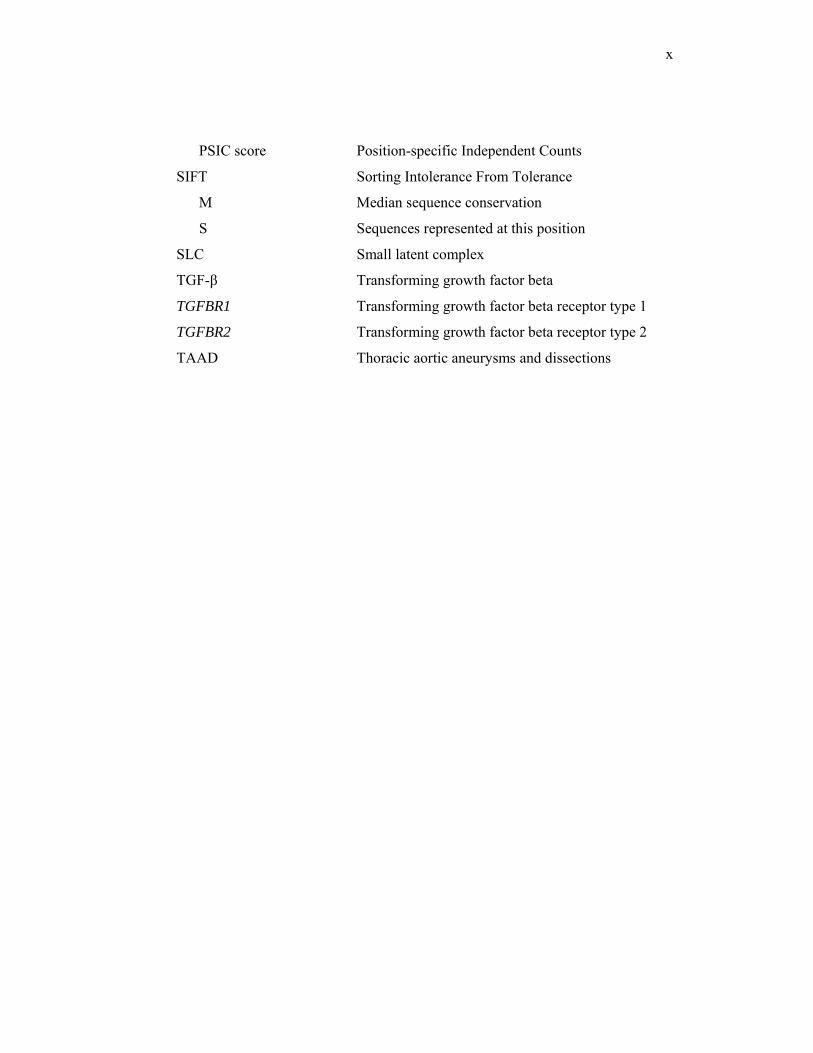
PSIC score Position-specific Independent Counts
SIFT Sorting Intolerance From Tolerance
M Median sequence conservation
S Sequences represented at this position
SLC Small latent complex
TGF-β Transforming growth factor beta
TGFBR1 Transforming growth factor beta receptor type 1
TGFBR2 Transforming growth factor beta receptor type 2
TAAD Thoracic aortic aneurysms and dissections
xi
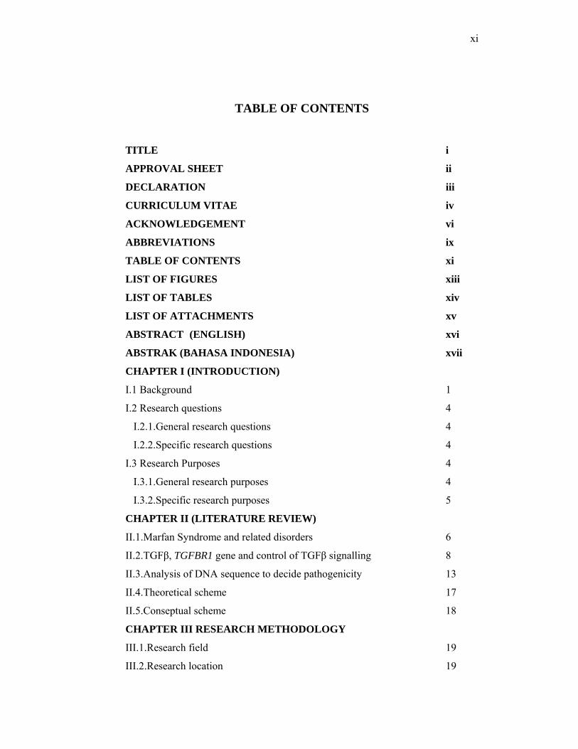
TABLE OF CONTENTS
TITLE i
APPROVAL SHEET ii
DECLARATION iii
CURRICULUM VITAE iv
ACKNOWLEDGEMENT vi
ABBREVIATIONS ix
TABLE OF CONTENTS xi
LIST OF FIGURES xiii
LIST OF TABLES xiv
LIST OF ATTACHMENTS xv
ABSTRACT (ENGLISH) xvi
ABSTRAK (BAHASA INDONESIA) xvii
CHAPTER I (INTRODUCTION)
I.1 Background 1
I.2 Research questions 4
I.2.1.General research questions 4
I.2.2.Specific research questions 4
I.3 Research Purposes 4
I.3.1.General research purposes 4
I.3.2.Specific research purposes 5
CHAPTER II (LITERATURE REVIEW)
II.1.Marfan Syndrome and related disorders 6
II.2.TGFβ, TGFBR1 gene and control of TGFβ signalling 8
II.3.Analysis of DNA sequence to decide pathogenicity 13
II.4.Theoretical scheme 17
II.5.Conseptual scheme 18
CHAPTER III RESEARCH METHODOLOGY
III.1.Research field 19
III.2.Research location 19
xii
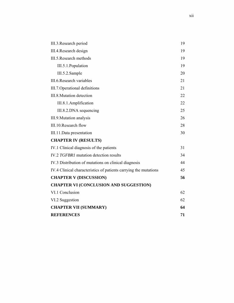
III.3.Research period 19
III.4.Research design 19
III.5.Research methods 19
III.5.1.Population 19
III.5.2.Sample 20
III.6.Research variables 21
III.7.Operational definitions 21
III.8.Mutation detection 22
III.8.1.Amplification 22
III.8.2.DNA sequencing 25
III.9.Mutation analysis 26
III.10.Research flow 28
III.11.Data presentation 30
CHAPTER IV (RESULTS)
IV.1 Clinical diagnosis of the patients 31
IV.2 TGFBR1 mutation detection results 34
IV.3 Distribution of mutations on clinical diagnosis 44
IV.4 Clinical characteristics of patients carrying the mutations 45
CHAPTER V (DISCUSSION) 56
CHAPTER VI (CONCLUSION AND SUGGESTION)
VI.1 Conclusion 62
VI.2 Suggestion 62
CHAPTER VII (SUMMARY) 64
REFERENCES 71

xiii
LIST OF FIGURES
Figure 1. Regulation of TGFβ bioavailability 8
Figure 2. Regulation of TGFβ bioavailability (cont.) 9
Figure 3. Signal transduction by TGFβ family members 10
Figure 4. Schematic diagram of TGFBR1 gene 11
Figure 5. Exons and domains organization 12
Figure 6. Bar graph showing the number of patients in each group 33
Figure 7. Mutation c.113G>A; p.C38Y in TGFBR1 (forward sequence) 36
Figure 8. Mutation c.451C>T; p.R151C in TGFBR1 (forward sequence) 37
Figure 9. Mutation c.605C>T; p.A202V in TGFBR1 (forward sequence) 37
Figure 10. Mutation c.839C>T; p.S280L in TGFBR1 (reverse sequence) 38
Figure 11. Mutation c.958A>G; p.I320V in TGFBR1 (forward sequence) 39
Figure 12. Mutation c.965G>A; p.G322D in TGFBR1 (forward sequence) 39
Figure 13. Mutation c.980C>T; p.P327L in TGFBR1 (forward sequence) 40
Figure 14. Mutation c.1282T>G; p.Y428D in TGFBR1 (forward sequence) 41
Figure 15. Mutation c.1460G>A; p.R487Q in TGFBR1 (reverse sequence) 42
Figure 16. Exons, domain organization and location of the mutations 42
Figure 17. Pedigree of patient 1 49
Figure 18. Pedigree of patient 3 50
Figure 19. Pedigree of patient 4 51
Figure 20. Pedigree of patient 8 52
Figure 21. Pedigree of patient 9 53
Figure 22. Pedigree of patient 10 54

xiv
LIST OF TABLES
Table 1. Clinical features of some overlapping disorders 7
Table 2. Primers sequence for amplifying the TGFBR1 gene exon 1-9 23
Table 3. M13 primers sequence 24
Table 4. Detail clinical features of MFS and related disorders patients based on
organ system presented in percentage 31
Table 5. Mutation, amino acid type changes and predicted functional effects of
amino acid substitution 35
Table 6. Multiple Sequence Alignment 43
Table 7. Polymorphisms found in this study 44
Table 8. Unclassified variants 46
Table 9. TGFBR1 mutations on clinical diagnosis 48
Table 10. Clinical findings of patient with TGFBR1 mutation 55

xv
LIST OF ATTACHMENTS Attachment 1. Ghent criteria of Marfan Syndrome 76
Attachment 2. Diagnostic criteria of some conditions overlapping with Marfan
Syndrome 79
Attachment 3. Diagnostic criteria of Aortic Aneurysms 83
Attachment 4. Laboratory Request form and Informed consent 85
Attachment 5. PolyPhen user guide 89
Attachment 6. SIFT user guide 97
xvi
ABSTRACT Background Marfan Syndrome (MFS) and related disorders involves particularly skeletal, ocular and cardiovascular. Aortic aneurysms and dissections is the commonest feature of MFS leading to death. MFS caused by mutation in FBN1, and recently, also in TGFBR2 and TGFBR1. Mutation analysis in TGFBR1 gene is needed to know if the mutation is present in patient with MFS and related disorders. Methods One hundred and ninety four patients with MFS and related disorders, who have at least one major criteria of MFS and found to be negative for FBN1 and TGFBR2 mutation, are included. The DNA of the patients were then analyzed for TGFBR1 mutation by direct sequencing of the whole gene. The potency of pathogenicity of the mutation was predicted by referring to previous publication, amino acid changes, multiple alignment analysis and with the help of internet-based software, PolyPhen and SIFT. Results Ten patients were found to carry TGFBR1 missense mutation. Each of them carried a different mutation, except 2 patients carried the same mutation. Seven out of nine of the mutations are considered pathogenic and 2 are not pathogenic. Aortic aneurysm is present in most patients with the mutation. None of the patient with classic MFS has mutation in TGFBR1 gene. Conclusion Despite of mutation analysis on FBN1 and TGFBR2, mutation analysis on TGFBR1 in patient with MFS and related disorders is needed, especially on those who have aortic aneurysm. Knowledge of the presence of a mutation in an individual or in a family, may give a better guidance for comprehensive treatment including genetic counseling
Keywords : Marfan Syndrome and related disorders, TGFBR1 mutation
xvii
ABSTRAK Latar Belakang Sindroma Marfan (MFS) dan kelainan terkait bermanifestasi di beberapa organ, terutama skeletal, okular dan kardiovaskular. Aneurysma dan diseksi aorta merupakan manifestasi yang paling sering mengakibatkan kematian pada MFS. MFS disebabkan oleh mutasi pada FBN1, dan akhir-akhir ini ditemukan juga disebabkan mutasi pada TGFBR2 dan TGFBR1. Analisis pada gen TGFBR1 diperlukan untuk mengetahui apakah pada pasien Marfan Syndrome dan kelainan terkait lainnya terdapat mutasi pada gen TGFBR1. Metode Sebanyak 194 pasien dengan MFS dan kelainan terkait yang memiliki paling tidak satu kelainan mayor diikutsertakan dalam penelitian ini. Sebelumnya, pasien telah terbukti tidak memiliki mutasi pada FBN1 dan TGFBR2. Sekuensing pada gen TGFBR1 dilakukan untuk mengetahui adanya mutasi. Potensi patogenisitas mutasi dianalisis dengan mengacu pada publikasi-publikasi sebelumnya, melihat perubahan asam amino, melakukan multiple alignment analysis dan menggunakan software PolyPhen dan SIFT. Hasil Didapatkan 10 pasien dengan mutasi pada TGFBR1, dari keseluruhan pasien yang diperiksa. Setiap pasien memiliki 1 missense mutation yang berbeda, kecuali 2 pasien dengan mutasi yang sama. Dari 9 missense mutations pada TGFBR1, 7 diantaranya patogenik dan 2 nonpatogenik. Aneurisma aorta merupakan manifestasi klinik yang muncul pada hampir semua pasien dengan mutasi. Mutasi pada TGFBR1 tidak ditemukan pada pasien dengan MFS klasik. Kesimpulan Analisis mutasi TGFBR1 pada MFS dan kelainan terkait tanpa mutasi di FBN1 dan TGFBR2 perlu dilakukan, terutama pada pasien dengan aneurisma aorta. Pengetahuan tentang keberadaan mutasi pada individu dalam keluarga dapat menjadi petujuk penting untuk penanganan yang komprehensif termasuk konseling genetika. Kata kunci : Sindroma Marfan dan kelainan terkait, mutasi TGFBR1
1
Chapter I
INTRODUCTION
I.1 Background
Marfan Syndrome (MFS), a common autosomal dominant inherited
disorder of fibrous connective tissue, has an estimated incidence of 1 :
5,000.1,2 This syndrome involves many organ systems, particularly the
skeletal, ocular and cardiovascular system. The most important life-
threatening complication in MFS is the occurrence of thoracic aortic
aneurysms leading to aortic dissection, rupture, or both.3
MFS is known to be one of the diseases in the spectrum of type-1
fibrillinopathies, which constitute a range of clinical phenotypes that are
caused by mutation in the gene for fibrillin-1 (FBN1 gene).1,2,4 In many cases,
a diagnosis of MFS can be established by the Ghent criteria.5 However, the
interpretation of these criteria is not always easy, due to the large clinical
range of fibrillinopathies that overlap with MFS, and to age-dependent
manifestations.
The initial idea from previous publications about the pathogenesis of
MFS concentrated on a static dominant negative model based on the concept
of fibrillin-rich micro fibrils as purely architectural elements in the extra
cellular matrix. Mutations in the fibrillin-1 gene (FBN1 gene), known to
cause MFS, however, have not always been found in MFS patients. Recent
2
findings of the pathogenesis of MFS demonstrate changes in growth factor
signaling and other changes in matrix-cell interactions.4
A connection of Marfan syndrome with the TGFβ signalling pathway
was initially found in a study on mouse model of Marfan Syndrome with
FBN1 mutation, and having lung emphysema as phenotypic manifestation.
This mouse model showed increased TGFβ signalling.6 The involvement of
TGFβ-receptor gene mutation in MFS has been shown in a Japanese patient
with MFS who had a balanced chromosomal translocation involving
chromosome 3p24. This locus had been found to show genetic linkage with
MFS in a large French pedigree. The breakpoint in the Japanese patient
disrupted the TGFBR2 gene. The same gene had a point mutation in the
French Marfan family.7 Later research on TGFβ showed that the use of TGFβ
antagonists such as TGFβ neutralizing antibody or the angiotensin II type 1
receptor blocker, Losartan, reduce the growth of aortic aneurysm in a mouse
model.8
The proteins fibrillin-1, TGFBR1 and TGFBR2 take part in
transforming growth factors β (TGFβ) signaling, thus mutations in one of
these gene could cause similar phenotypes. TGFβ is stored in the extra
cellular matrix in a latent form, bound to fibrillin 1 to form a complex. The
complex is released by proteases, and the active TGFβ binds to its receptors
on the cell surface (TGFβR1 and TGFβR2), leading to dimerization of the
receptor. The kinase domain of the receptor is then activated and starting a
signaling cascade in the cell regulating a number of cellular processes such as
3
apoptosis, inflammation, proliferation and growth.9 Thus, TGFβ signaling
will depend on the amount of latent TGFβ present in the tissue, strength of
the binding of the complex and activity of TGFβ receptors.
Mutations in the TGFBR1 and TGFBR2 genes have also been reported
in individuals with Loeys-Dietz aortic aneurysms syndrome, a syndrome
characterized by hypertelorism, bifid uvula and/or cleft palate, generalized
arterial tortuosity with ascending aortic aneurysm, and worse cardiovascular
risk profile than classic MFS.10 Another study reported TGFBR1 and
TGFBR2 mutations in individuals with MFS-like phenotypes who previously
tested negative for mutations in FBN1 gene.11 Mutations in TGFBR1 have
been found in other syndromes related with MFS, e.g. Sphrintzen-Goldberg
Syndrome, and in patients with Thoracic Aortic Aneurysms and Dissection
(TAAD).6,11,12 So far, in total 22 different mutations have been found in the
TGFBR1 gene.13 The phenotypes of patients having the mutations in TGFBR
genes could not be clearly differentiated from each other.
In this descriptive research we looked for and analyzed mutations in
the TGFBR1 gene in patients referred to the DNA laboratory of Vrije
Universiteit Medisch Centrum Amsterdam (VUmc), The Netherlands, with a
clinical suspicion of MFS or related disorders, who did not have a FBN1 or
TGFBR2 mutation.

4
I.2 Research Questions
I.2.1 General research question :
What kind of mutations can be found in the TGFBR1 gene in
people with clinical Marfan Syndrome, and other related disorders with
negative FBN1 and TGFBR2 mutations?
I.2.2 Specific research question
1. Is there any mutation in the TGFBR1 gene as a candidate gene for
Marfan Syndrome and related disorders with negative FBN1 and
TGFBR2 mutations, and if yes, what kind of mutation is it?
2. How is the prediction of pathogenicity of the mutation?
3. How is the distribution of clinical phenotype on genotype?
4. Is there any clinical characteristic that may lead to TGFBR1 gene
mutation analysis?
I.3 Research purposes
I.3.1 General purposes :
To identify and analyze the kind of mutations in the TGFBR1 gene
as candidate gene for Marfan Syndrome and related disorders with
negative FBN1 and TGFBR2 mutations, and to see the distribution of
clinical phenotype on the genotype .
5
I.3.2 Specific purposes :
1. To detect the mutations in the TGFBR1 gene in a person with Marfan
Syndrome and related disorders with negative FBN1 and TGFBR2
mutations.
2. To analyze the kind of mutations and the potential pathogenic effect
of the mutations.
3. To see the distribution of clinical phenotype on the genotype.
4. To see whether there is a clinical characteristic that may lead to
TGFBR1 mutation analysis.
6
Chapter II
LITERATURE REVIEW
II.1 MARFAN SYNDROME AND RELATED DISORDERS
Patients with Marfan Syndrome (MFS) may have abnormalities in
several different organ systems, but mostly in skeletal, ocular and
cardiovascular systems.1 Skeletal features of MFS are increased height,
disproportionately long limbs and digits, elbow contracture, anterior chest
deformity, mild to moderate joint laxity, vertebral column deformity (scoliosis
and thoracic lordosis) and a narrow, high palate with crowding of the teeth.
Ocular findings in MFS include increased axial globe length, corneal flatness
and (sub) luxation of the lenses (ectopia lentis). Mitral valve prolaps, mitral
regurgitation, dilatation of the aortic root and aortic regurgitation are
cardiovascular features. Aneurysm of the aorta and aortic dissection are the
major life-threatening cardiovascular complications. Mostly, this feature
brings MFS into special attention. Other common features are striae distensae,
pulmonary blebs, which predispose to spontaneous pneumothorax and spinal
arachnoid cysts or diverticula. By CT or MRI scanning also dural ectasia can
be found. The early-onset severe MFS, neonatal MFS, presents with serious
cardiovascular abnormalities as well as congenital contractures. MFS is also
associated with a high prevalence of obstructive sleep apneu.1,2,14,15
The diagnosis of MFS is based on a set of clinical diagnostic criteria,
termed The Ghent Criteria.5 In clinical practice, these criteria are not always
7
obvious, since there are many conditions overlapping with MFS and because
of age-dependent manifestation. The overlapping conditions are Familial
Aortic Aneurysm, Bicuspid Aortic Valve with Aortic Dilatation, Familial
Ectopia Lentis, MASS phenotype, Marfan Body Type, Mitral Valve Prolapse
Syndrome, Congenital Contractural Arachnodactily (Beals syndrome),
Stickler syndrome, Shprintzen-Goldberg Syndrome, Loeys-Dietz Syndrome
and Ehlers-Danlos syndrome.1,2,4,14,15 The clinical features of those overlap
disorders are described in the table below :
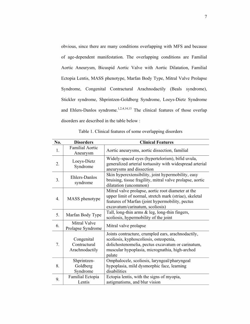
Table 1. Clinical features of some overlapping disorders
No. Disorders Clinical Features
1. Familial Aortic Aneurysm Aortic aneurysms, aortic dissection, familial
2. Loeys-Dietz Syndrome
Widely-spaced eyes (hypertelorism), bifid uvula, generalized arterial tortuosity with widespread arterial aneurysms and dissection
3. Ehlers-Danlos syndrome
Skin hyperextensibility, joint hypermobility, easy bruising, tissue fragility, mitral valve prolapse, aortic dilatation (uncommon)
4. MASS phenotype
Mitral valve prolapse, aortic root diameter at the upper limit of normal, stretch mark (striae), skeletal features of Marfan (joint hypermobility, pectus excavatum/carinatum, scoliosis)
5. Marfan Body Type Tall, long-thin arms & leg, long-thin fingers, scoliosis, hypermobility of the joint
6. Mitral Valve Prolapse Syndrome Mitral valve prolapse
7. Congenital
Contractural Arachnodactily
Joints contracture, crumpled ears, arachnodactily, scoliosis, kyphoscoliosis, osteopenia, dolichostenomelia, pectus excavatum or carinatum, muscular hypoplasia, micrognathia, high-arched palate
8. Shprintzen-Goldberg Syndrome
Omphalocele, scoliosis, laryngeal/pharyngeal hypoplasia, mild dysmorphic face, learning disabilities
9. Familial Ectopia Lentis
Ectopia lentis, with the signs of myopia, astigmatisms, and blur vision
8
This table shows some of the disorders that have overlapping phenotypes
with Marfan Syndrome.
II.2 TGFβR1, TGFBR1 GENE AND CONTROL OF TGFβ SIGNALLING
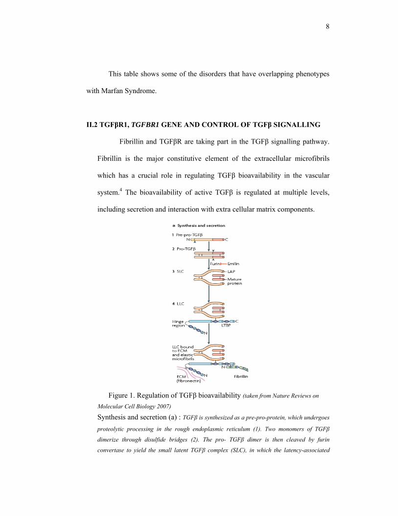
Fibrillin and TGFβR are taking part in the TGFβ signalling pathway.
Fibrillin is the major constitutive element of the extracellular microfibrils
which has a crucial role in regulating TGFβ bioavailability in the vascular
system.4 The bioavailability of active TGFβ is regulated at multiple levels,
including secretion and interaction with extra cellular matrix components.
Figure 1. Regulation of TGFβ bioavailability (taken from Nature Reviews on
Molecular Cell Biology 2007)
Synthesis and secretion (a) : TGFβ is synthesized as a pre-pro-protein, which undergoes
proteolytic processing in the rough endoplasmic reticulum (1). Two monomers of TGFβ
dimerize through disulfide bridges (2). The pro- TGFβ dimer is then cleaved by furin
convertase to yield the small latent TGFβ complex (SLC), in which the latency-associated
9
peptide (LAP) and the mature peptide are connected (3). This processing step is inhibited by
emilin-1. The large latent TGFβ binding protein (LTBP) is attached, and form the large latent
TGFβ complex (LLC) (4). The N-terminal and hinge region of LTBP interact covalently with
extra cellular matrix component, such as fibronectin. The C-terminal region of LTBP interacts
non covalently with the N-terminal region of fibrillin-1.9
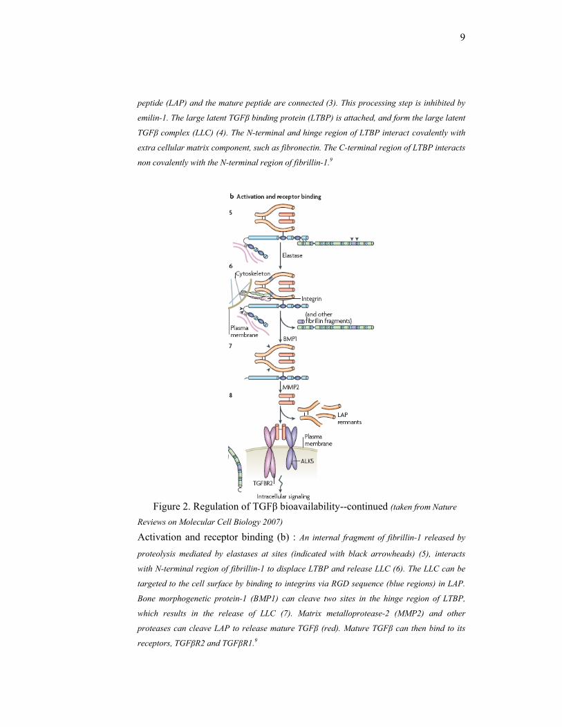
Figure 2. Regulation of TGFβ bioavailability--continued (taken from Nature
Reviews on Molecular Cell Biology 2007)
Activation and receptor binding (b) : An internal fragment of fibrillin-1 released by
proteolysis mediated by elastases at sites (indicated with black arrowheads) (5), interacts
with N-terminal region of fibrillin-1 to displace LTBP and release LLC (6). The LLC can be
targeted to the cell surface by binding to integrins via RGD sequence (blue regions) in LAP.
Bone morphogenetic protein-1 (BMP1) can cleave two sites in the hinge region of LTBP,
which results in the release of LLC (7). Matrix metalloprotease-2 (MMP2) and other
proteases can cleave LAP to release mature TGFβ (red). Mature TGFβ can then bind to its
receptors, TGFβR2 and TGFβR1.9
10
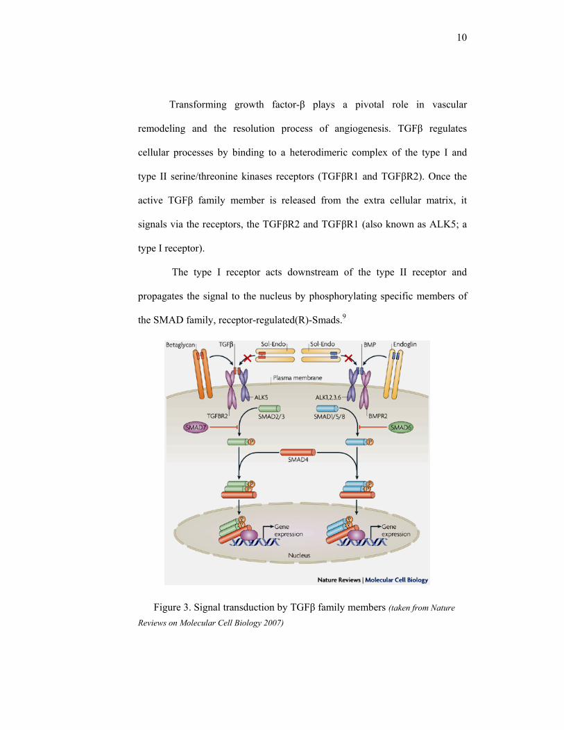
Transforming growth factor-β plays a pivotal role in vascular
remodeling and the resolution process of angiogenesis. TGFβ regulates
cellular processes by binding to a heterodimeric complex of the type I and
type II serine/threonine kinases receptors (TGFβR1 and TGFβR2). Once the
active TGFβ family member is released from the extra cellular matrix, it
signals via the receptors, the TGFβR2 and TGFβR1 (also known as ALK5; a
type I receptor).
The type I receptor acts downstream of the type II receptor and
propagates the signal to the nucleus by phosphorylating specific members of
the SMAD family, receptor-regulated(R)-Smads.9
Figure 3. Signal transduction by TGFβ family members (taken from Nature
Reviews on Molecular Cell Biology 2007)
11
The type I receptor acts downstream of the type II receptor and
propagates the signal to the nucleus by phosphorylating specific members of
the SMAD family, receptor-regulated(R)-Smads.9 The phosphorylated
SMADs will then gives signal to the nucleus, and regulates the transcription
steps of the genes which play roles in differentiation, growth inhibition,
deposition of extra cellular matrix and apoptosis.
TGFβR1 (ALK5) is required for TGFβ-ALK1 activation, whereas
ALK1 inhibits intracellular ALK5-SMAD signaling. The differential
activation of these two distinct type-I receptor pathways by TGFβ provides the
endothelial cells with an intricate mechanism to precisely regulate, and even
switch between, TGFβ-induced biological responses. For example, TGFβ-
ALK1 activation leads to stimulation of endothelial cell proliferation and
migration, whereas TGFβ-ALK5 activation inhibits these responses.9
The TGFBR1 gene is also known as activin A receptor like kinase, or
serine/threonine-protein kinase receptor R4 gene. The DNA size is
approximately 45kb long, the mRNA size is 2308bp, contains of 9 exons and
is located on chromosome 9q22.33.16,17 The schematic diagram of The
TGFBR1 gene with its exons and introns is presented in the figure below :
contains of 9 exons and is located on chromosome 9q22.33.16,17
12
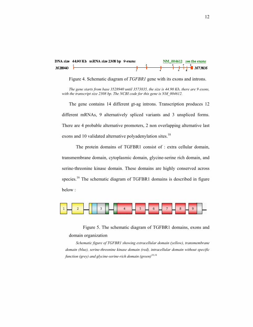
Figure 4. Schematic diagram of TGFBR1 gene with its exons and introns.
The gene starts from base 3528940 until 3573835, the size is 44.90 Kb, there are 9 exons, with the transcript size 2308 bp. The NCBI code for this gene is NM_004612.
The gene contains 14 different gt-ag introns. Transcription produces 12
different mRNAs, 9 alternatively spliced variants and 3 unspliced forms.
There are 4 probable alternative promoters, 2 non overlapping alternative last
exons and 10 validated alternative polyadenylation sites.18
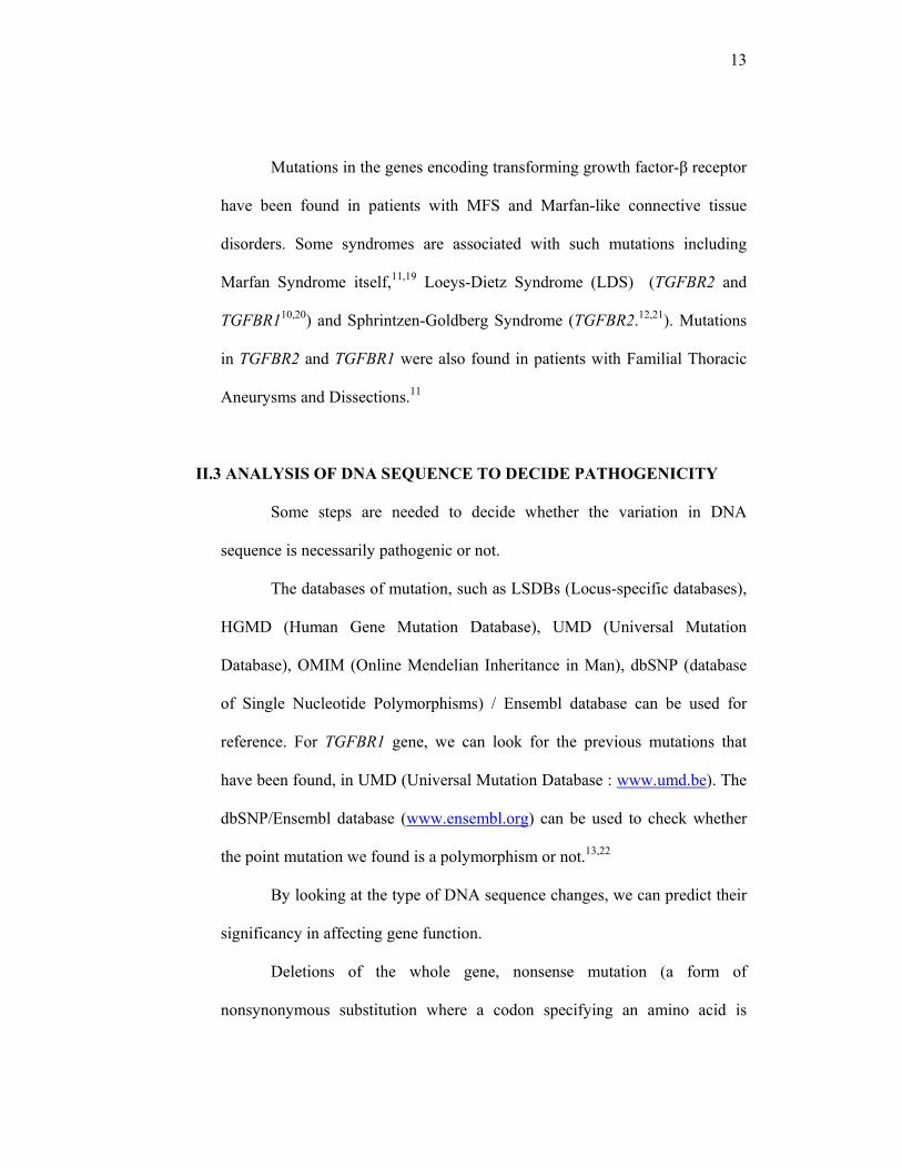
The protein domains of TGFBR1 consist of : extra cellular domain,
transmembrane domain, cytoplasmic domain, glycine-serine rich domain, and
serine-threonine kinase domain. These domains are highly conserved across
species.16 The schematic diagram of TGFBR1 domains is described in figure
below :
Figure 5. The schematic diagram of TGFBR1 domains, exons and
domain organization Schematic figure of TGFBR1 showing extracellular domain (yellow), transmembrane
domain (blue), serine-threonine kinase domain (red), intracellular domain without specific
function (grey) and glycine-serine-rich domain (green)10,16
13
Mutations in the genes encoding transforming growth factor-β receptor
have been found in patients with MFS and Marfan-like connective tissue
disorders. Some syndromes are associated with such mutations including
Marfan Syndrome itself,11,19 Loeys-Dietz Syndrome (LDS) (TGFBR2 and
TGFBR110,20) and Sphrintzen-Goldberg Syndrome (TGFBR2.12,21). Mutations
in TGFBR2 and TGFBR1 were also found in patients with Familial Thoracic
Aneurysms and Dissections.11
II.3 ANALYSIS OF DNA SEQUENCE TO DECIDE PATHOGENICITY
Some steps are needed to decide whether the variation in DNA
sequence is necessarily pathogenic or not.
The databases of mutation, such as LSDBs (Locus-specific databases),
HGMD (Human Gene Mutation Database), UMD (Universal Mutation
Database), OMIM (Online Mendelian Inheritance in Man), dbSNP (database
of Single Nucleotide Polymorphisms) / Ensembl database can be used for
reference. For TGFBR1 gene, we can look for the previous mutations that
have been found, in UMD (Universal Mutation Database : www.umd.be). The
dbSNP/Ensembl database (www.ensembl.org) can be used to check whether
the point mutation we found is a polymorphism or not.13,22
By looking at the type of DNA sequence changes, we can predict their
significancy in affecting gene function.
Deletions of the whole gene, nonsense mutation (a form of
nonsynonymous substitution where a codon specifying an amino acid is
14
replaced by a stop codon) and frameshift mutation (a mutation that alters the
normal translational reading frame of an mRNA by adding or deleting a
number of bases that is not a multiple of three), are almost certain to destroy
gene function.23
Mutation that change the conserved splice site (GT…AG nucleotides)
affects splicing, and will usually abolish the function of the gene. In silico
predictions for splice site are available, for example Splice Sequence Finder
(Montpelier) www.umd.be/SSF, GeneSplicer Web Interface
www.tigr.org/tdb/GeneSplicer/gene_spl.html, etc.23
A missense mutation is more likely to be pathogenic if it affects a part
of the protein domain known to be functionally important.23
Changing of an amino acid is more likely to affect function if that
amino acid is conserved in related genes, orthologs (genes present in different
genomes which are directly related through descent from a common ancestor)
or paralogs (genes present in a single genome as a result of gene duplication).
If two or more sequences show sufficient degree of similarity (sequence
homology), they can be assumed to be derived from the same ancestor. The
higher the degree of similarity, the gene are more conserved, means that the
gene has very important role through evolution. The mutation at that point will
be strongly suspicious to be pathogenic. Multiple alignment analysis is
comparing the amino acid sequence of certain protein with the closest similar
sequences from some species. By looking at the position and the presence of
15
amino acid, we can decide whether the amino acid is conserved across the
species or not.23,24
Amino acid substitutions are more likely to affect function if they are
nonconservative. Nonconservative substitutions result in replacement of one
amino acid by another that is chemically not similar. For example, the change
from a polar to a non polar amino acid, or an acidic to a basic.23
Another way to predict the potential pathogenicity of a mutation is by
using in silico prediction analysis. There are some software available in the
internet, that can be used to do the prediction, for example PolyPhen
(Polymorphisms Phenotyping) and SIFT (Sorting Intolerance From
Tolerance). PolyPhen (http://coot.embl.de/PolyPhen/)25, is an automatic tool
for prediction of possible impact of an amino acid substitution on the structure
and function of human protein. This prediction is based on empirical rules
which are applied to the sequence, phylogenetic and structural information
characterizing the substitution. A protein identifier from proteins database,
such as SWALL is needed before entering the amino acid substitution. This
program will then identify the sites in which the new amino acid replaced, do
multiple alignment, and calculate the so-called profile matrix by Position-
Specific Independent Counts (PSIC). The PSIC score will be used as one of
prediction parameter. A Protein Quarternary Structure (PQS) database is also
used as another consideration. The results of PolyPhen can be : probably
damaging (it is with high confidence supposed to affect protein function or
structure), possibly damaging (supposed to affect protein function or
16
structure), benign (most likely lacking any phenotypic effect) and unknown (in
some rare cases, when the lack of data do not allow PolyPhen to make a
prediction).26 The detail guideline to interpreting PolyPhen result is attached in
the attachment.
SIFT BLink is a sequence-homology-based tool that sorts intolerant
from tolerant amino acid substitutions and predict whether an amino acid
substitution at a particular position in a protein will have a phenotypic effects.
SIFT BLink bases its prediction on sequence data alone and does not depend
on knowledge of protein structure and function. The results of SIFT BLink
prediction are affect protein function and tolerated (means that the substitution
can be tolerated, thus does not affect protein function). The sequence data for
specific protein is inputted, and will be followed by some steps in which SIFT
BLink process the data input to prediction. Substitutions at each position with
normalized probabilities less than a chosen cutoff are predicted to be
deleterious, while those greater than or equal to the cutoff are predicted to be
tolerated.24
17
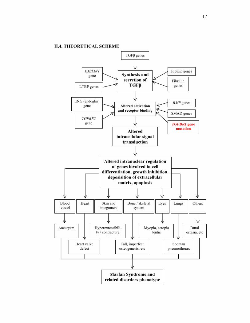
II.4. THEORETICAL SCHEME
TGFβ genes
Altered intracellular signal
transduction
Synthesis and secretion of
TGFβ
Altered activation and receptor binding
LTBP genes
EMILIN1gene
Fibrillin genes
Fibulin genes
TGFBR2 gene
ENG (endoglin) gene
TGFBR1 gene mutation
BMP genes
SMAD genes
Aneurysm Hyperextensibili- ty / contracture,
etc
Myopia, ectopia lentis
Heart valve defect
Tall, imperfect osteogenesis, etc
Spontan pneumothorax
Dural ectasia, etc
Blood vessel
Heart Skin and integumen
Bone / skeletal system
Eyes Lungs Others
Altered intranuclear regulation of genes involved in cell
differentiation, growth inhibition, deposisition of extracellular
matrix, apoptosis
Marfan Syndrome and related disorders phenotype

18
Notes : - TGFβ = Transforming growth factor beta - LTBP = Latent transforming growth factor binding protein - BMP = Bone morphogenetic protein - TGFBR1 = Transforming growth factor beta receptor type 1 - TGFBR2 = Transforming growth factor beta receptor type 2
II.5 CONCEPTUAL SCHEME
The conseptual scheme of this research
TGFBR1 gene
Mutations at any specific
sites
Marfan Syndrome and related disorders
phenotypes
19
Chapter III
RESEARCH METHODOLOGY
III.1. Research field
This research is in the field of medical genetics.
III.2. Research location
This research was held in the DNA Diagnostic Laboratory of Vrije
Universiteit Medisch Centrum (VUmc), Amsterdam, The Netherlands for DNA
analysis.
III.3. Research period
This research has been conducted in one year.
III.4. Research design
This is a descriptive study.
III.5. Research methods
III.5.1. Population
The population of this research is the DNA samples of patients
with Marfan Syndrome and related disorders which have been reffered to
DNA Diagnostic Laboratory of VUmc Hospital Amsterdam, The
Netherlands from the year 1998-2008.
20
III.5.2. Samples
The DNA samples were donation with permission from Gerard
Pals, PhD as the principal investigator of Connective Tissue Disorders
research in the DNA Diagnostic Laboratory of VUmc Hospital Amsterdam,
The Netherlands. All of the samples used in this research are part of
Connective Tissue Disorders research project, and have been consent to be
included in research (informed consent form attached).
We selected the first 194 unrelated patient’s from VUmc’s DNA
Diagnostic Laboratory database by their registration numbers, which have
been referred as Marfan Syndrome, suspected Marfan Syndrome, or related
disorders. The phenotypic characteristics of the patients were then traced
from their laboratory request form.
III.5.2.1. Inclusion criteria :
1. Having at least one major criterion of MFS
2. Found to be negative for FBN1 and TGFBR2 mutations.
III.5.2.2. Exclusion criteria :
1. Not enough amount of DNA available for complete
examination.
III.5.2.3. Minimum sample requirement :
This is the first research on TGFBR1 gene in Marfan Syndrome
and related disorders patients in The Netherlands. Another
research on TGFBR1 revealed a frequency of 4% among
numbers of patients.19 Sample amount determination for
estimation of proportion in population is as below 28 :

21
P=0.04; Zα= 1.96; d=0.10 n= (1.96)2 x 0.04 x (1-0.04) = 153 (0.10) 2 Notes :
P = the proportion of TGFBR1 mutations found in previous study = 0.0419
d = precision level = 0.10 α = significancy level = 0.95, Zα = 1.96 The minimum sample which is required is 153 samples.
III.6. Research Variables :
The variables of this research are :
1. Clinical phenotypes of Marfan Syndrome and related disorders
Scale : nominal
2. Mutation in TGFBR1 gene
Scale : nominal
3. Pathogenicity of mutation
Scale : nominal
III.7. Operational Definitions
1. Marfan Syndrome : a group of clinical signs, fulfilling the Revised
Criteria of Marfan Syndrome (Ghent Criteria).
2. Ghent Criteria of Marfan Syndrome : clinical criteria for diagnosing
Marfan Syndrome (details attached in the attachment). For the index
cases, major criteria in at least 2 different organ systems and
involvement in third organ is needed, if the family/genetic history is
not contributory. For a relative of an index case, one major criterion in
n = Zα2 PQ d2
22
an organ system and an involvement of second organ is needed if a
major criterion in family history is present.
3. Suspected MFS : incomplete Ghent criteria with more than one signs
which are mentioned in the criteria.
4. Related disorders of Marfan Syndrome : disorders that share several
symptoms with Marfan Syndrome, including Loeys-Dietz Syndrome,
Ehler-Danlos Syndrome vascular type, Aortic aneurysms and
dissection, Bicuspid Aortic Valve with Aortic Dilatation, Familial
Ectopia Lentis, MASS phenotype, Mitral Valve Prolapse Syndrome,
Congenital Contractural Arachnodactily (Beals syndrome), Stickler
syndrome, Shprintzen-Goldberg Syndrome, joint hypermobility, etc.
The clinical features of these disorders are in the attachment.
5. Phenotype : all the clinical signs found in Marfan Syndrome and
related disorders patients
6. Mutation : an alteration in DNA sequence.
7. Pathogenicity : the condition in which the mutation will results in
protein changes thus causing disease, predicted by the type of amino
acid changes, domain localization, multiple alignment, and prediction
results of internet-based software, with consideration to literature.
III.8. Mutation Detection
III.8.1 Amplification
In order to get enough amount of DNA fragment to be visible in
the gel and have strong enough signal in sequencing, the TGFBR1
23
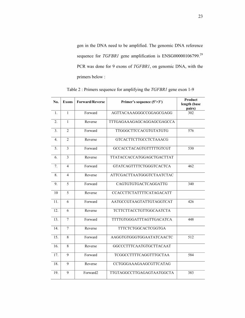
gen in the DNA need to be amplified. The genomic DNA reference
sequence for TGFBR1 gene amplification is ENSG00000106799.29
PCR was done for 9 exons of TGFBR1, on genomic DNA, with the
primers below :
Table 2 : Primers sequence for amplifying the TGFBR1 gene exon 1-9
No. Exons Forward/Reverse Primer’s sequence (5’>3’) Product length (base
pairs) 1. 1 Forward AGTTACAAAGGGCCGGAGCGAGG 302
2. 1 Reverse TTTGAGAAAGAGCAGGAGCGAGCCA
3. 2 Forward TTGGGCTTCCACGTGTATGTG 576
4. 2 Reverse GTCACTTCTTGCCTCTAAACG
5. 3 Forward GCCACCTACAGTGTTTTTGTCGT 530
6. 3 Reverse TTATACCACCATGGAGCTGACTTAT
7. 4 Forward GTATCAGTTTTCTGGGTCACTCA 462
8. 4 Reverse ATTCGACTTAATGGGTCTAATCTAC
9. 5 Forward CAGTGTGTGACTCAGGATTG 340
10 5 Reverse CCACCTTCTATTTTCATAGACATT
11. 6 Forward AATGCCGTAAGTATTGTAGGTCAT 426
12. 6 Reverse TCTTCTTACCTGTTGGCAATCTA
13. 7 Forward TTTTGTGGGATTTAGTTGACATCA 448
14. 7 Reverse TTTCTCTGGCACTCGGTGA
15. 8 Forward AAGGTGTGGGTGGAATATCAACTC 512
16. 8 Reverse GGCCCTTTCAATGTGCTTACAAT
17. 9 Forward TCGGCCTTTTCAGGTTTGCTAA 584
18. 9 Reverse CCTGGGAAAGAAGCGTTCATAG
19. 9 Forward2 TTGTAGGCCTTGAGAGTAATGGCTA 383

24
Table shows the sequence of each primer (forward and reverse) which is used to amplified exon 1 to 9 of TGFBR1 gene, and the product size.
Notes : For exon 9 we used 2 forward primers, because of a long T-
stretch in the DNA sequence. A long T-strech is vulnerable to deletion, so
that the sequence output will be messy, and the mutation after the deleted-T
will not be detected. The other forward primer which start after T-stretch
will prevent the undetected mutation.
At the 5’ end of each primer an M13 tail primer sequence forward or
reverse (M13 primer, INVITROGEN, Cat.No.N520-02 (F) and N530-02
(R)) was added, in order to simplify the sequencing procedure. With M13
tail primer attached in the PCR primer, the amplified fragment will start
from M13 sequence, so that in cycle-sequencing reaction we will need only
M13 tail primer to amplify all exon, and not different primer for different
exons. The sequences of M13 tails primers are as below :
Table 3 : M13 primers sequence
No. Primer’s name Sequences
1. M13 forward GTAAAACGACGGCCAG
2. M13 reverse CAGGAAACAGCTATGA
The table shows the sequences of M13 tail primer, which is attached to PCR primer and used in cycle-sequencing reaction.
Five microliter DNA solution (DNA concentration : 20 ng/µl) was
added into 25 µl PCR mixture, which contained 0.2 µl of 25 mM dNTPs,
0.75 µl of 50 mM MgCl2 (Invitrogen), 1µl of 10pmol/µl each primer
(Invitrogen), 2.5 µl of 10x PCR buffer (Invitrogen), 0.2 µl of 5U/µl Taq
DNA polymerase (Platinum Taq DNA Polymerase, Invitrogen,
25
Cat.No.10966-034) and 15.35 µl H2O. The thermal profile included initial
denaturation for 5 minutes at 940C, followed by 35 cycles of denaturation (1
minute at 940C), annealing (1 minute at 650C), and extension (1 minute at
720C), in PE9700 Applied Biosystem thermocycler. Five microliters of each
sample was then runned on an 2% agarose gel with 100V for 30 minutes and
stained with ethidium bromide, to confirm PCR amplification product (the
size of PCR product as described in table 1).
III.8.2 DNA sequencing
The purpose of sequencing is to determine the order of the nucleotides
of a gene.
Prior to sequencing, the PCR products were purified from excess
primers and dNTPs molecule by a mixture of Exo 1 (Exonuclease 1, USB
Corp. Cleveland, Ohio, Cat.No.70073X) and SAP enzyme (Shrimp Alkaline
Phosphatase, USB Corp. Cleveland, Ohio, Cat.No.70092Y). Five microliters
of PCR were taken into the reaction, together with 0.25 µL SAP, 0.25 µL
Exo1 and 1.5 µL HPLC H2O. The mixture was then incubated in a
thermocycler with a program of 30 minutes in 370C followed by 15 minutes
in 800C. Then diluted with 15 µL HPLC H2O, and was added sequencing
primer (forward or reverse) as much as 1 µL.
Sequencing reactions used the BigDye Terminator Cycle Sequencing
kit (version 3 Applied Biosystems. Foster City, CA, USA, Cat.No.4737458)
on an ABI 3730 Genetic Analyzer (Applied Biosystems, Foster City, Ca,

26
USA). Seven microliter of BigDye mix, which contained of 0.5 µL BigDye
V3.1 reaction mix, 1.75 µL BigDye V3.1 5x sequencing buffer and 4.75 µL
HPLC H2O, were taken into reaction together with 3 µL of SAP-Exo1-PCR
product mixture. Then runned in a thermocycler with a program of 960C 10
minutes denaturation, 550C 5 seconds annealing, 600C 4 minutes elongation,
25 cycles.
The products of sequencing reaction were then precipitated using
ethanol precipitation method in order to remove unincorporated dye
terminators. The product would then be added with 20 µL formamide,
heated in thermocycler on 940C for 2 minutes and cooled down to 40C, and
put in the sequencer (ABI 3730 Genetic Analyzer, Applied Biosystem)
III.9. Mutation Analysis
We compared the sequence of patients with the reference sequence.
The variant numbering is based on the cDNA sequence
(ENST00000374994),30 where +1 corresponds to the nucleotide A of ATG,
the translation initial codon.
The UMD database of TGFBR1 mutations, the Ensemble SNPs
database of polymorphisms and the previous reports on TGFBR1, were used
to confirm the DNA sequence variants. Whenever the variant was not
mentioned as polymorphism in one of those references, we did the analysis
based on the changes in amino acid types, domain conservation in some
27
species, protein structure and previous publications on the mutations.
Internet-based software programs to predict the possible impact of amino
acid substitutions were also used to help the analysis.
The first program was PolyPhen (http://coot.embl.de/PolyPhen/)25, a
web-based tool to predict the possible impact of amino acid substitution on
the structure and function of the protein. The data query needs a protein
identifier which codes specific protein in the protein database. The protein
identifier in SWALL-protein database for TGFBR1 is P36897.31 We use
default query parameters for protein quaternary structure (PQS) databases
and performing calculations for all hits. The second program we used was
SIFT Blink27, a sequence homology-based amino acid substitution
prediction method (available at
http://blocks.fhcrc.org/sift/SIFT_BLink_submit.html). We applied
gi:4759226 protein sequence of TGFBR1 by using parameter “best BLAST
hit to each organism” and omitting sequences 100% identical to query.
Results were reported as “affects protein function” or “tolerated” according
to this analysis.
To help predict the affect of mutation on the splice site, we used web-
based tool Human Splicing Finder32 (available at www.umd.be/HSF/) which
analyzed the sequence towards the presence of enhancer motifs, silencer
motifs, exonic splicing regulatory sequences, potential branch points and
potential splice sites.
28
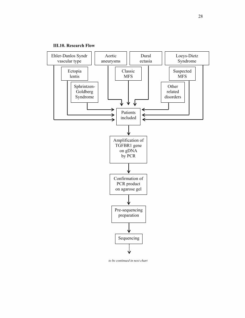
III.10. Research Flow
to be continued in next chart
Classic MFS
Loeys-Dietz Syndrome
Ehler-Danlos Syndr vascular type
Sphrintzen-Goldberg Syndrome
Aortic aneurysms
Dural ectasia
Ectopia lentis
Other related
disorders
Suspected MFS
Patients included
Amplification of TGFBR1 gene
on gDNA by PCR
Sequencing
Confirmation of PCR product
on agarose gel
Pre-sequencing preparation
29
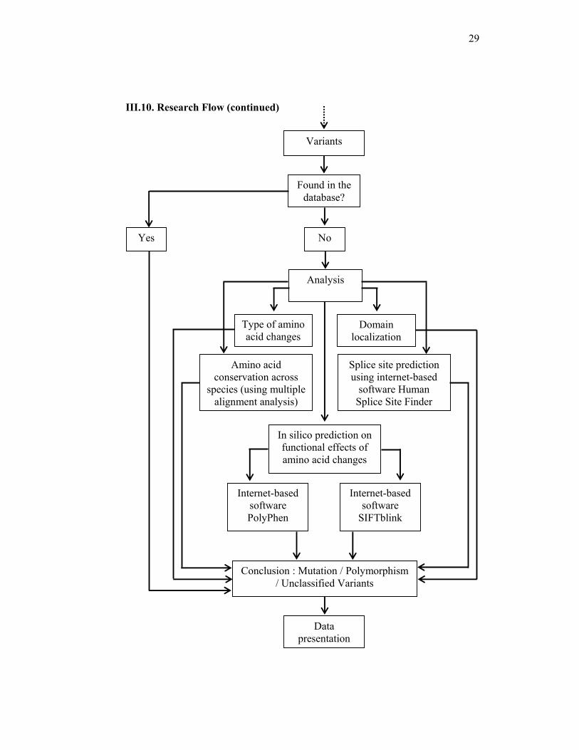
III.10. Research Flow (continued)
Analysis
Type of amino acid changes
Domain localization
Amino acid conservation across
species (using multiple alignment analysis)
In silico prediction on functional effects of amino acid changes
Internet-based software PolyPhen
Internet-based software
SIFTblink
Splice site prediction using internet-based
software Human Splice Site Finder
Conclusion : Mutation / Polymorphism / Unclassified Variants
Variants
Found in the database?
Yes No
Data presentation

30
III.11. Data Analysis
The data will be analyzed descriptively for the clinical features of
the patients, the number of patients in each diagnosis group, the mutations that
have been found and the distribution in each exon and domain, the amino acid
type changes and the prediction of pathogenicity with their multiple sequence
alignment, and the polymorphisms and unclassified variants. The details are as
below :
1. The clinical features of the patients; the list of mutations, amino acid-
type changes and the prediction results from PolyPhen and SIFT; the
list of polymorphisms and unclassified variants and the distribution of
TGFBR1 mutations on clinical diagnosis will be presented in tables.
2. The number of patients in each diagnosis will be presented in graph.
3. The distribution of mutations in each exon and domain will be
presented in schematic figure.
4. The multiple sequence alignment will be presented in figure.
31
Chapter IV
RESULTS
IV.1 Clinical diagnosis of patients
The patient samples included in this study came from many centers,
inside and outside The Netherlands, such as Belgium and United Kingdom.
All the DNA samples included are a donation with permission from DNA
diagnostic laboratory of Vrije Universiteit Amsterdam, The Netherlands.
The clinical information of patients described here has been collected
from clinical observations that have been mentioned in the laboratory
request form.
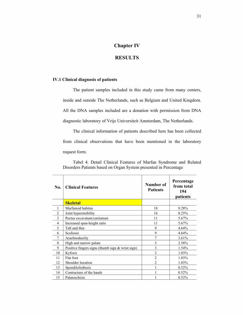
Tabel 4. Detail Clinical Features of Marfan Syndrome and Related Disorders Patients based on Organ System presented in Percentage
No. Clinical Features Number of Patients
Percentage from total
194 patients
Skeletal 1 Marfanoid habitus 18 9.28% 2 Joint hypermobility 16 8.25% 3 Pectus excavatum/carinatum 11 5.67% 4 Increased span-height ratio 11 5.67% 5 Tall and thin 9 4.64% 6 Scoliosis 9 4.64% 7 Arachnodactily 7 3.61% 8 High and narrow palate 5 2.58% 9 Positive fingers signs (thumb sign & wrist sign) 3 1.54% 10 Kyfosis 2 1.03% 11 Flat foot 2 1.03% 12 Shoulder luxation 2 1.03% 13 Spondilolisthesis 1 0.52% 14 Contracture of the hands 1 0.52% 15 Palatoschizis 1 0.52%

32
No. Clinical Features Number of Patients
Percentage from total
194 patients
16 Crowded teeth 1 0.52% 17 Skeletal abnormalities (unspecified) 26 13.40%
Cardiovascular 1 Aortic aneurysms 137 70.62% 2 Aortic dissection 24 12.37% 3 Mitral Valve Prolaps 5 2.58% 4 Aortic valve insufficiency 4 2.06% 5 Pulmonary stenosis 2 1.03% 6 Dissections of artery coronaria 2 1.03% 7 Aneurysms of other big vessel 1 0.52% 8 Persisten Ductus Arteriosus 1 0.52% 9 Mitral Insufficiency 1 0.52% 10 Varices 1 0.52% 11 Heart problem (unspecified) 1 0.52%
Occular 1 Myopia 4 2.06% 2 Lens subluxation 4 2.06% 3 Ectopia Lentis 3 1.54% 4 Flat cornea 1 0.52% 5 Retinal detachment 1 0.52% 6 Eye abnormality (unspecified) 7 3.61%
Lung 1 Spontaneous pneumothorax 3 1.54% 2 Lung abnormality (unspecified) 2 1.03%
Dura 1 Dural ectasia 6 3.09%
Skin & Integumen 1 Striae 5 2.58% 2 Thin skin 2 1.03% 3 Uterus & Bladder prolaps 2 1.03% 4 Hernia inguinalis 1 0.52% 5 Skin abnormality unspecified 4 2.06%
Others 1 Uvula bifida 2 1.03% 2 Mental retardation 1 0.52%
Notes : one patient may have more than one clinical features.
The clinical diagnoses of the patients were based on clinical findings
and matched with Ghent Criteria. A diagnosis of MFS was based on Ghent
33
Criteria. Incomplete Ghent Criteria, or having at least one major criterion in
an organ system with minor criterion of another organ, or more than one
minor criterion, would be considered as Suspected MFS. The patients with
only specific clinical features (such as only has aortic aneurysm, ectopia
lentis, dural ectasia or joint hypermobility) would be grouped as the clinical
findings, recognized as Marfan Syndrome, Suspected MFS, Aortic
Aneurysms and/ Dissections, Familial Aortic Aneurysms and/ Dissections,
Ectopia Lentis, Dural Ectasia, Joint Hypermobility.
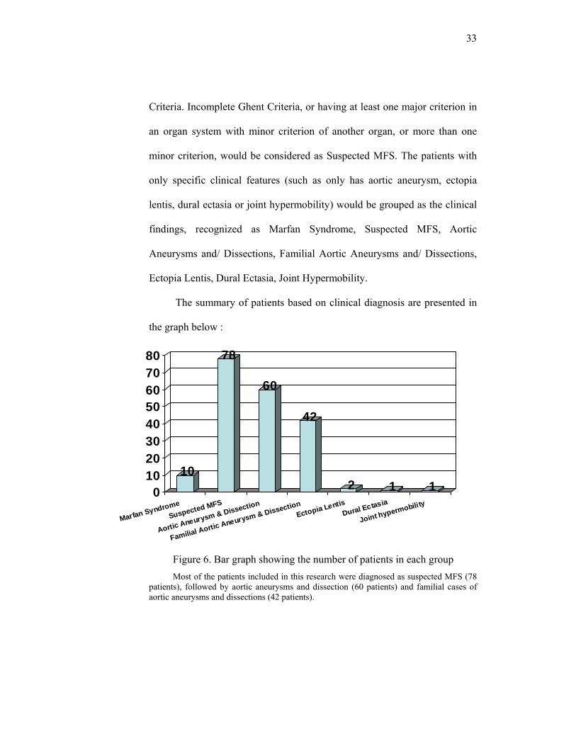
The summary of patients based on clinical diagnosis are presented in
the graph below :
Figure 6. Bar graph showing the number of patients in each group Most of the patients included in this research were diagnosed as suspected MFS (78
patients), followed by aortic aneurysms and dissection (60 patients) and familial cases of aortic aneurysms and dissections (42 patients).
10
78
60
42
2 1 101020304050607080
Marfan SyndromeSuspected MFS
Aortic Aneurysm & Dissection
Familial Aortic Aneurysm & DissectionEctopia Lentis
Dural Ectasia
Joint hypermobility

34
IV.2 TGFBR1 mutation detection results
On sequencing all 9 exons of TGFBR1, a total of 9 mutations, 7
different polymorphisms and 3 unclassified variants in TGFBR1 were found.
The mutations were found in 10 patients. The 9 mutations, occured in 7
different exons (see table 5).
We did analysis on mutations by observing the amino acid changes,
looking at the conservation in 11 different species and the domain
localization, and using internet-based software to predict the pathogenicity
of amino acid changes.
The list of mutations, amino acid-type changes and the prediction
results from PolyPhen and SIFT are presented in table 3 below :
35
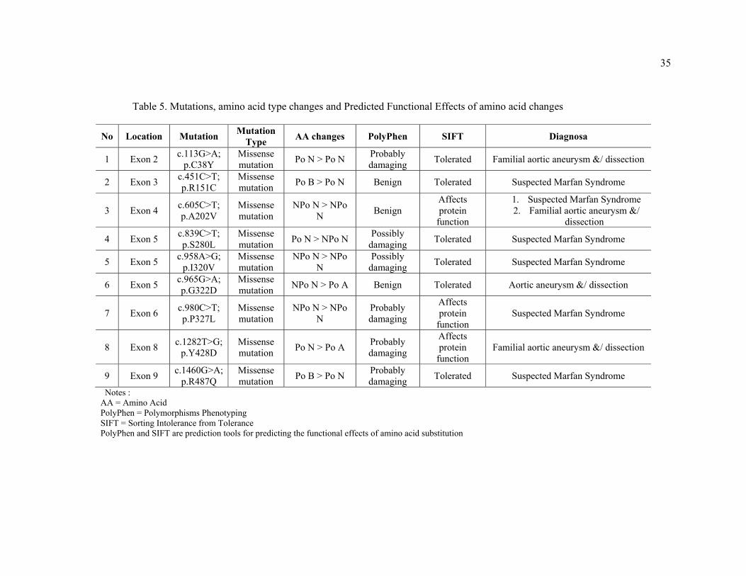
Table 5. Mutations, amino acid type changes and Predicted Functional Effects of amino acid changes
No Location Mutation Mutation Type AA changes PolyPhen SIFT Diagnosa
1 Exon 2 c.113G>A; p.C38Y
Missense mutation Po N > Po N Probably
damaging Tolerated Familial aortic aneurysm &/ dissection
2 Exon 3 c.451C>T; p.R151C
Missense mutation Po B > Po N Benign Tolerated Suspected Marfan Syndrome
3 Exon 4 c.605C>T; p.A202V
Missense mutation
NPo N > NPo N Benign
Affects protein
function
1. Suspected Marfan Syndrome 2. Familial aortic aneurysm &/
dissection
4 Exon 5 c.839C>T; p.S280L
Missense mutation Po N > NPo N Possibly
damaging Tolerated Suspected Marfan Syndrome
5 Exon 5 c.958A>G; p.I320V
Missense mutation
NPo N > NPo N
Possibly damaging Tolerated Suspected Marfan Syndrome
6 Exon 5 c.965G>A; p.G322D
Missense mutation NPo N > Po A Benign Tolerated Aortic aneurysm &/ dissection
7 Exon 6 c.980C>T; p.P327L
Missense mutation
NPo N > NPo N
Probably damaging
Affects protein
function Suspected Marfan Syndrome
8 Exon 8 c.1282T>G; p.Y428D
Missense mutation Po N > Po A Probably
damaging
Affects protein
function Familial aortic aneurysm &/ dissection
9 Exon 9 c.1460G>A; p.R487Q
Missense mutation Po B > Po N Probably
damaging Tolerated Suspected Marfan Syndrome
Notes : AA = Amino Acid PolyPhen = Polymorphisms Phenotyping SIFT = Sorting Intolerance from Tolerance PolyPhen and SIFT are prediction tools for predicting the functional effects of amino acid substitution

36
F H
F
C
F Y H
Explanation of the table and sequencing results :
All of the mutations are missense mutations, in which a nucleotide substitution
results in an amino acid change :
1. The mutation is located in exon 2 of TGFBR1 gene, at the position 113 of
cDNA, in which guanine is replaced by adenine, resulted in the change of
amino acid 38 from cysteine (a polar-neutral amino acid) to tyrosine (a
polar-neutral). This mutation is predicted to be probably damaging by
PolyPhen and tolerated by SIFT.
The position of mutation in gene sequence is shown below :
Figure 7. Mutation c.113G>A; p.C38Y in TGFBR1 (forward sequence) Mutation in exon 2, showed a Cysteine (TGC) change to Tyrosine (TAC).
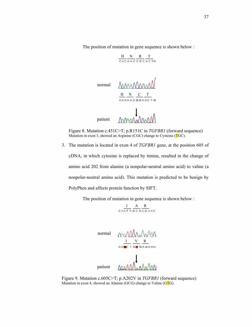
2. The mutation is located in exon 3 of TGFBR1 gene, at the position 451 of
cDNA, in which cytosine is replaced by timine, resulted in the change of
amino acid 151 from arginine (a polar-basic amino acid) to cysteine (a
polar-neutral amino acid). This mutation is predicted to be benign by
PolyPhen and tolerated by SIFT.
normal
patient
37
normal
H N R T
H N C T
I
R
A R
I V
The position of mutation in gene sequence is shown below :
Figure 8. Mutation c.451C>T; p.R151C in TGFBR1 (forward sequence) Mutation in exon 3, showed an Arginine (CGC) change to Cysteine (TGC).
3. The mutation is located in exon 4 of TGFBR1 gene, at the position 605 of
cDNA, in which cytosine is replaced by timine, resulted in the change of
amino acid 202 from alanine (a nonpolar-neutral amino acid) to valine (a
nonpolar-neutral amino acid). This mutation is predicted to be benign by
PolyPhen and affects protein function by SIFT.
The position of mutation in gene sequence is shown below :
Figure 9. Mutation c.605C>T; p.A202V in TGFBR1 (forward sequence) Mutation in exon 4, showed an Alanine (GCG) change to Valine (GTG).
normal
patient
patient
38
V S D
V L D
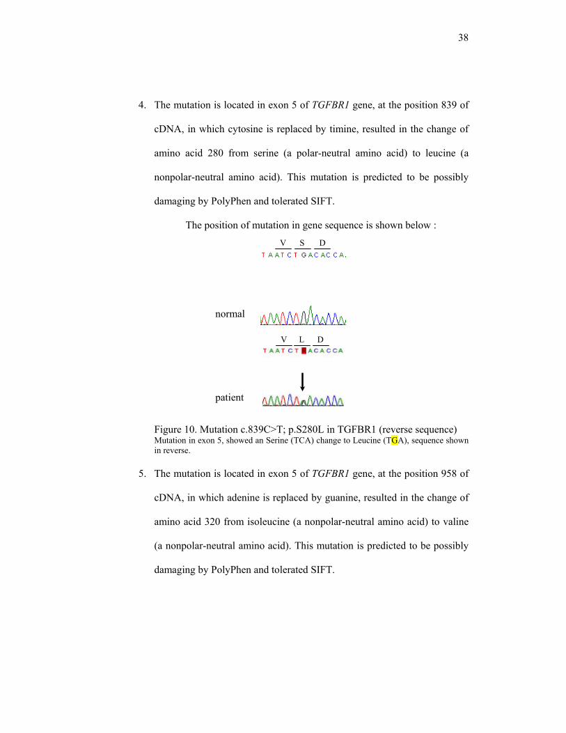
4. The mutation is located in exon 5 of TGFBR1 gene, at the position 839 of
cDNA, in which cytosine is replaced by timine, resulted in the change of
amino acid 280 from serine (a polar-neutral amino acid) to leucine (a
nonpolar-neutral amino acid). This mutation is predicted to be possibly
damaging by PolyPhen and tolerated SIFT.
The position of mutation in gene sequence is shown below :
Figure 10. Mutation c.839C>T; p.S280L in TGFBR1 (reverse sequence) Mutation in exon 5, showed an Serine (TCA) change to Leucine (TGA), sequence shown in reverse.
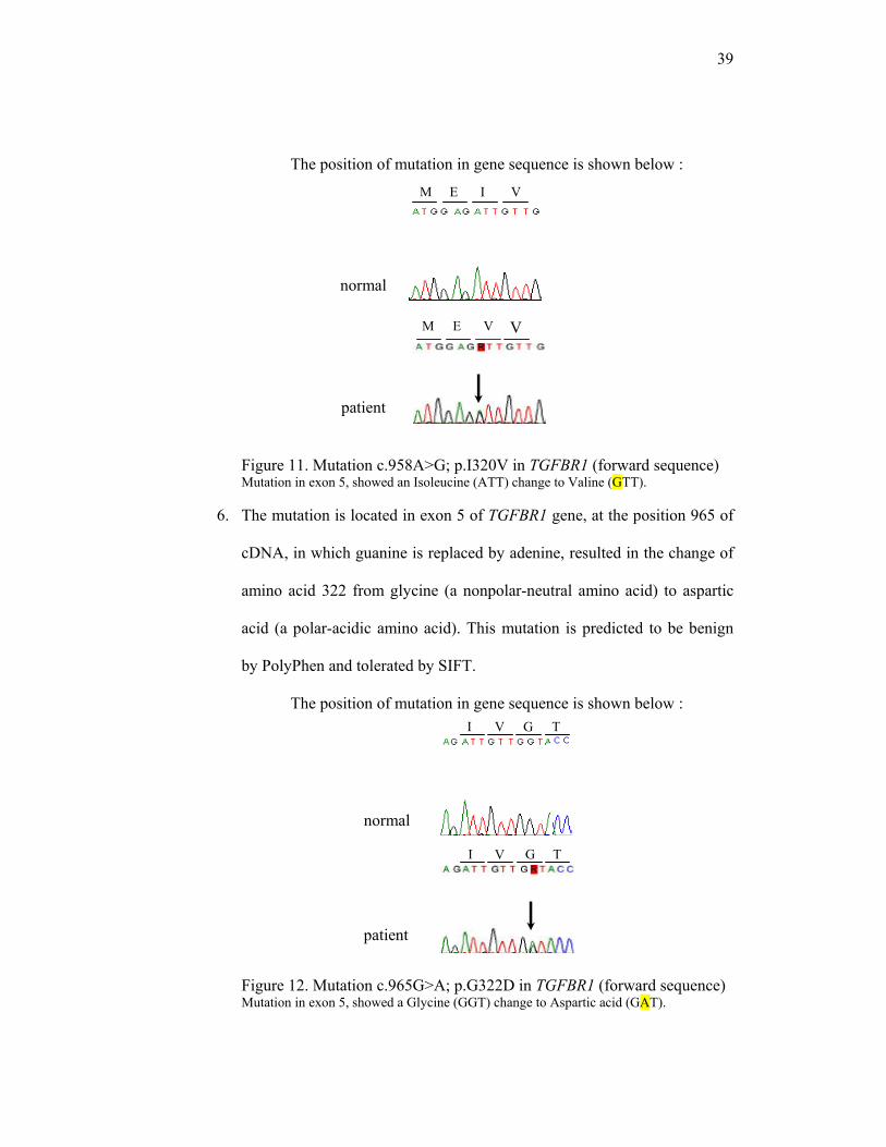
5. The mutation is located in exon 5 of TGFBR1 gene, at the position 958 of
cDNA, in which adenine is replaced by guanine, resulted in the change of
amino acid 320 from isoleucine (a nonpolar-neutral amino acid) to valine
(a nonpolar-neutral amino acid). This mutation is predicted to be possibly
damaging by PolyPhen and tolerated SIFT.
normal
patient
39
patient
M E I V
M E I V
I V G T
V T G I
The position of mutation in gene sequence is shown below :
Figure 11. Mutation c.958A>G; p.I320V in TGFBR1 (forward sequence) Mutation in exon 5, showed an Isoleucine (ATT) change to Valine (GTT).
6. The mutation is located in exon 5 of TGFBR1 gene, at the position 965 of
cDNA, in which guanine is replaced by adenine, resulted in the change of
amino acid 322 from glycine (a nonpolar-neutral amino acid) to aspartic
acid (a polar-acidic amino acid). This mutation is predicted to be benign
by PolyPhen and tolerated by SIFT.
The position of mutation in gene sequence is shown below :
Figure 12. Mutation c.965G>A; p.G322D in TGFBR1 (forward sequence) Mutation in exon 5, showed a Glycine (GGT) change to Aspartic acid (GAT).
normal
V
patient
normal
40
A L K
P A K
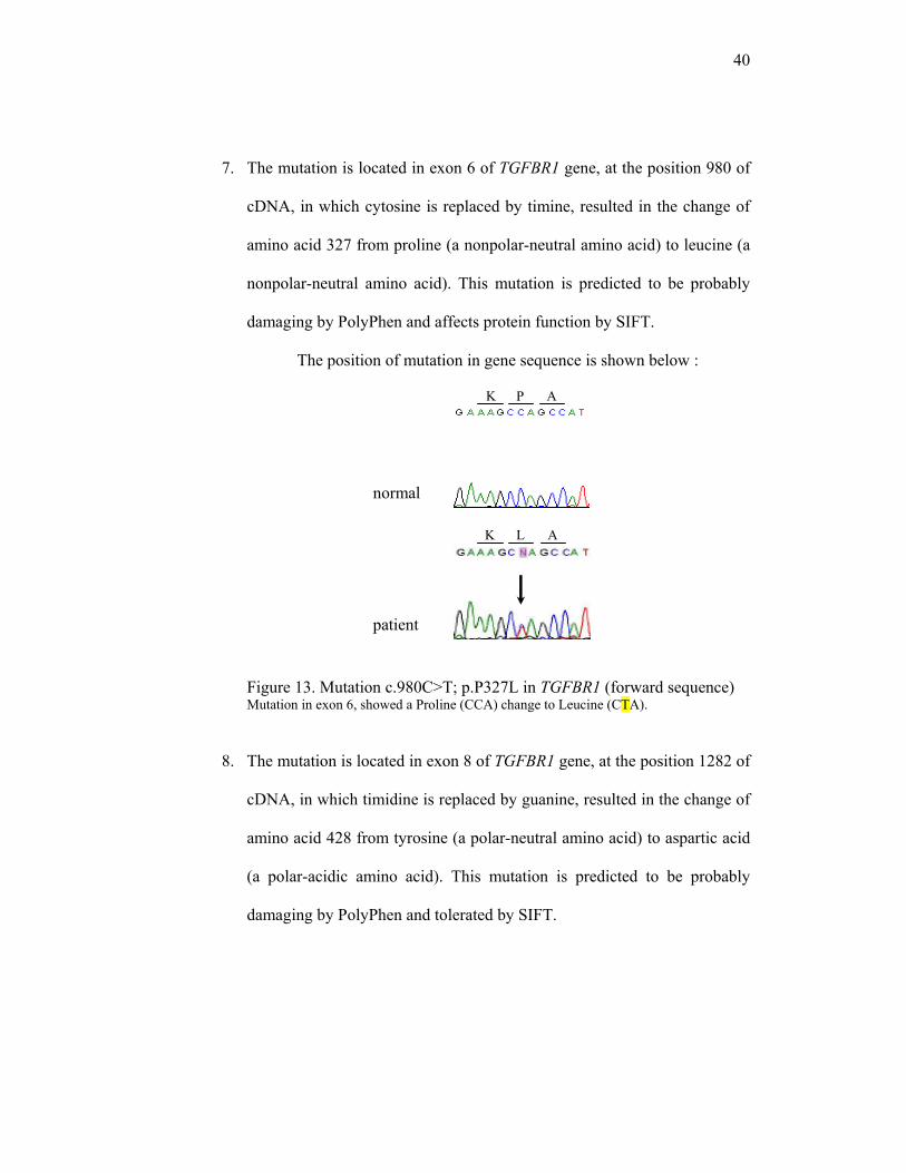
7. The mutation is located in exon 6 of TGFBR1 gene, at the position 980 of
cDNA, in which cytosine is replaced by timine, resulted in the change of
amino acid 327 from proline (a nonpolar-neutral amino acid) to leucine (a
nonpolar-neutral amino acid). This mutation is predicted to be probably
damaging by PolyPhen and affects protein function by SIFT.
The position of mutation in gene sequence is shown below :
Figure 13. Mutation c.980C>T; p.P327L in TGFBR1 (forward sequence) Mutation in exon 6, showed a Proline (CCA) change to Leucine (CTA).
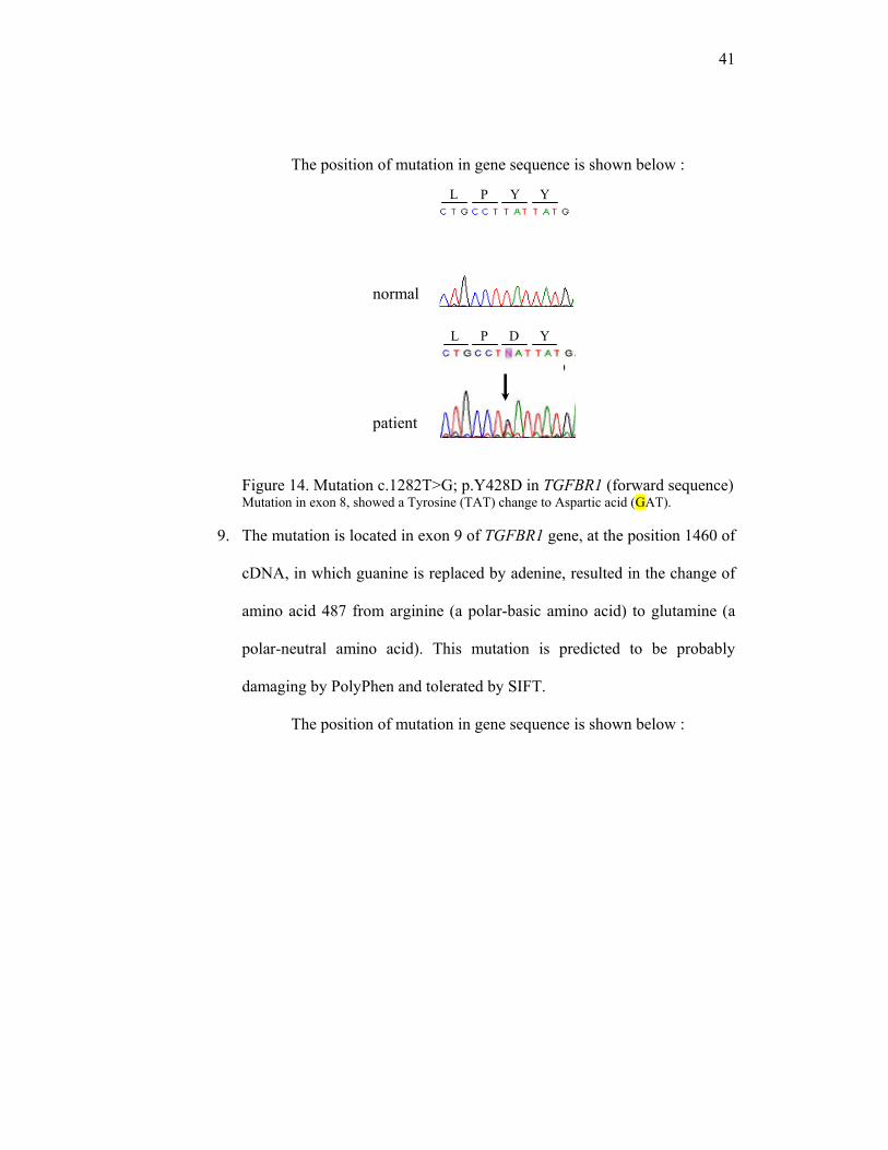
8. The mutation is located in exon 8 of TGFBR1 gene, at the position 1282 of
cDNA, in which timidine is replaced by guanine, resulted in the change of
amino acid 428 from tyrosine (a polar-neutral amino acid) to aspartic acid
(a polar-acidic amino acid). This mutation is predicted to be probably
damaging by PolyPhen and tolerated by SIFT.
patient
normal
41
Y D P L
Y Y P L
The position of mutation in gene sequence is shown below :
Figure 14. Mutation c.1282T>G; p.Y428D in TGFBR1 (forward sequence) Mutation in exon 8, showed a Tyrosine (TAT) change to Aspartic acid (GAT).
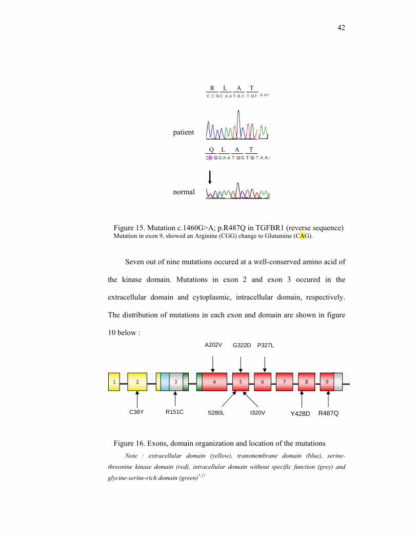
9. The mutation is located in exon 9 of TGFBR1 gene, at the position 1460 of
cDNA, in which guanine is replaced by adenine, resulted in the change of
amino acid 487 from arginine (a polar-basic amino acid) to glutamine (a
polar-neutral amino acid). This mutation is predicted to be probably
damaging by PolyPhen and tolerated by SIFT.
The position of mutation in gene sequence is shown below :
normal
patient
42
normal
T A L Q
T A L R
Figure 15. Mutation c.1460G>A; p.R487Q in TGFBR1 (reverse sequence) Mutation in exon 9, showed an Arginine (CGG) change to Glutamine (CAG).
Seven out of nine mutations occured at a well-conserved amino acid of
the kinase domain. Mutations in exon 2 and exon 3 occured in the
extracellular domain and cytoplasmic, intracellular domain, respectively.
The distribution of mutations in each exon and domain are shown in figure
10 below :
Figure 16. Exons, domain organization and location of the mutations Note : extracellular domain (yellow), transmembrane domain (blue), serine-
threonine kinase domain (red), intracellular domain without specific function (grey) and
glycine-serine-rich domain (green)7,17
C38Y Y428D R487QR151C S280L I320V
P327L G322D A202V
patient
43
From the picture above we can see that most of the mutations are located in
exon 5 (3 out of 9 different mutations).
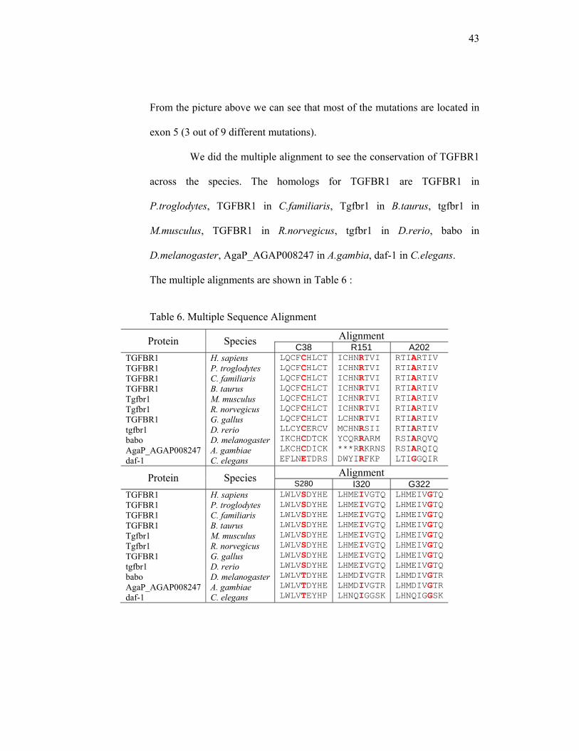
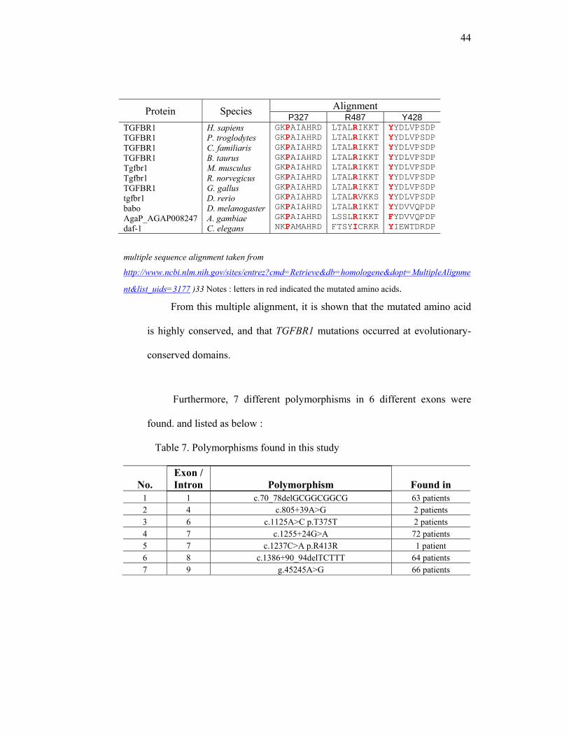
We did the multiple alignment to see the conservation of TGFBR1
across the species. The homologs for TGFBR1 are TGFBR1 in
P.troglodytes, TGFBR1 in C.familiaris, Tgfbr1 in B.taurus, tgfbr1 in
M.musculus, TGFBR1 in R.norvegicus, tgfbr1 in D.rerio, babo in
D.melanogaster, AgaP_AGAP008247 in A.gambia, daf-1 in C.elegans.
The multiple alignments are shown in Table 6 :
Table 6. Multiple Sequence Alignment
Protein Species Alignment C38 R151 A202
TGFBR1 TGFBR1 TGFBR1 TGFBR1 Tgfbr1 Tgfbr1 TGFBR1 tgfbr1 babo AgaP_AGAP008247 daf-1
H. sapiens P. troglodytes C. familiaris B. taurus M. musculus R. norvegicus G. gallus D. rerio D. melanogasterA. gambiae C. elegans
LQCFCHLCTLQCFCHLCTLQCFCHLCTLQCFCHLCT LQCFCHLCT LQCFCHLCT LQCFCHLCT LLCYCERCVIKCHCDTCKLKCHCDICKEFLNETDRS
ICHNRTVI ICHNRTVI ICHNRTVI ICHNRTVI ICHNRTVI ICHNRTVI LCHNRTVI MCHNRSII YCQRRARM ***RRKRNSDWYIRFKP
RTIARTIV RTIARTIV RTIARTIV RTIARTIV RTIARTIV RTIARTIV RTIARTIV RTIARTIV RSIARQVQ RSIARQIQ LTIGGQIR
Protein Species Alignment S280 I320 G322
TGFBR1 TGFBR1 TGFBR1 TGFBR1 Tgfbr1 Tgfbr1 TGFBR1 tgfbr1 babo AgaP_AGAP008247 daf-1
H. sapiens P. troglodytes C. familiaris B. taurus M. musculus R. norvegicus G. gallus D. rerio D. melanogasterA. gambiae C. elegans
LWLVSDYHELWLVSDYHELWLVSDYHELWLVSDYHELWLVSDYHELWLVSDYHELWLVSDYHELWLVSDYHELWLVTDYHELWLVTDYHELWLVTEYHP
LHMEIVGTQLHMEIVGTQLHMEIVGTQLHMEIVGTQLHMEIVGTQLHMEIVGTQLHMEIVGTQLHMEIVGTQ LHMDIVGTRLHMDIVGTRLHNQIGGSK
LHMEIVGTQ LHMEIVGTQ LHMEIVGTQ LHMEIVGTQ LHMEIVGTQ LHMEIVGTQ LHMEIVGTQ LHMEIVGTQ LHMDIVGTR LHMDIVGTR LHNQIGGSK
44
Protein Species Alignment P327 R487 Y428
TGFBR1 TGFBR1 TGFBR1 TGFBR1 Tgfbr1 Tgfbr1 TGFBR1 tgfbr1 babo AgaP_AGAP008247 daf-1
H. sapiens P. troglodytes C. familiaris B. taurus M. musculus R. norvegicus G. gallus D. rerio D. melanogasterA. gambiae C. elegans
GKPAIAHRDGKPAIAHRDGKPAIAHRDGKPAIAHRDGKPAIAHRDGKPAIAHRDGKPAIAHRDGKPAIAHRDGKPAIAHRDGKPAIAHRDNKPAMAHRD
LTALRIKKTLTALRIKKT LTALRIKKT LTALRIKKT LTALRIKKT LTALRIKKT LTALRIKKT LTALRVKKS LTALRIKKTLSSLRIKKTFTSYICRKR
YYDLVPSDP YYDLVPSDP YYDLVPSDP YYDLVPSDP YYDLVPSDP YYDLVPSDP YYDLVPSDP YYDLVPSDP YYDVVQPDP FYDVVQPDP YIEWTDRDP
multiple sequence alignment taken from
http://www.ncbi.nlm.nih.gov/sites/entrez?cmd=Retrieve&db=homologene&dopt=MultipleAlignme
nt&list_uids=3177 )33 Notes : letters in red indicated the mutated amino acids.
From this multiple alignment, it is shown that the mutated amino acid
is highly conserved, and that TGFBR1 mutations occurred at evolutionary-
conserved domains.
Furthermore, 7 different polymorphisms in 6 different exons were
found. and listed as below :
Table 7. Polymorphisms found in this study
No. Exon / Intron Polymorphism Found in
1 1 c.70_78delGCGGCGGCG 63 patients 2 4 c.805+39A>G 2 patients 3 6 c.1125A>C p.T375T 2 patients 4 7 c.1255+24G>A 72 patients 5 7 c.1237C>A p.R413R 1 patient 6 8 c.1386+90_94delTCTTT 64 patients 7 9 g.45245A>G 66 patients

45
Explanation of the table, starts from the first polymorphism :
1. Polymorphism 1 is located in exon 1, it is an in-frame deletion of 9
bases starts from position 70 of cDNA until 78 (GCGGCGGCG), and
causes deletion of 3 amino acid Alanin.
2. Polymorphism 2 is located in intron 4, in the position of cDNA
805+39, where adenine is replaced by guanine.
3. Polymorphism 3 is located in exon 6, in the position of cDNA 1125,
where adenine is replaced by cytosine. It is a silent mutation, because
the nucleotide change results in the same amino acid, Threonine.
4. Polymorphism 4 is located in intron 7, in the position of cDNA
1255+24 where guanine is replaced by adenine.
5. Polymorphism 5 is located in exon 7, in the position of cDNA 1237,
where cytosine is replaced by adenine. It is a silent mutation, because
the nucleotide change results in the same amino acid, Arginine.
6. Polymorphism 6 is located in intron 8. It is a deletion of 5 bases
(TCTTT) in the position of cDNA 1386+90 to 1386+94.
7. Polymorphism 7 is located in intron 9, in the position of gDNA 45245,
where adenine is replaced by guanine.
These 7 polymorphisms have been previously reported in the Ensembl
database of polymorphisms.29
Three variants are left as unclassified (see table 5). They neither have
been reported as polymorphisms, nor as mutations.

46
Table 8. Unclassified Variants (UV)
No. Exon / Intron
Unclassified Variants Found in Clinical Features
1 2 c.343+46T>G 1 patient Skeletal abnormality and aortic aneurysm
2 7 c.1255+103G>A 4 patients
1. Joint hypermobility, aortic aneurysm, pneumothorax 2. Familial aortic aneurysm 3. Scoliosis, pectus excavatum, arachnodactily, narrow & high palate 4. Pectus carinatum, flexible shoulder, tall & skinny
3 8 c.1386+87_91delTTTTC 1 patient Joint hypermobility, aortic aneurysm
Explanation of the table, starts from the first UV :
1. UV 1 is located in intron 2, in the position of cDNA 343+46, where
timidine is replaced by guanine. This UV presents in patient with
skeletal abnormality and aortic aneurysm.
2. UV 2 is located in intron 7, in the position of cDNA 1255+103, where
guanine is replaced by adenine. This UV presents in 4 patients with :
1. Joint hypermobility, aortic aneurysm and pneumothorax.
2. Familial aortic aneurysm.
3. Scoliosis, pectus excavatum, arachnodactily, narrow and high
palate.
4. Pectus carinatum, flexible shoulder, tall and skinny.
3. UV 3 is located in intron 8. It is a deletion of 5 bases (TTTTC), starts
from c.1386+87 to 1386+91. This UV presents in patient with joint
hypermobility and aortic aneurysm.

47
All of the Uvs are non-coding variants (located in intron, which are not
code the amino acid). To decide the pathogenicity, they need to be analyzed
on cDNA to see whether this UV affecting splice site, therefore tissue
biopsies of these patients are needed to perform the analyses. The DNA of
parents are unfortunately unavailable.
48
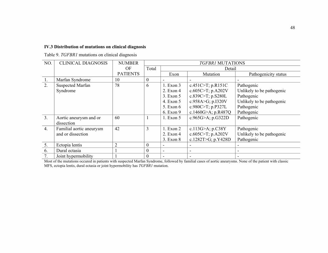
IV.3 Distribution of mutations on clinical diagnosis
Table 9. TGFBR1 mutations on clinical diagnosis
NO. CLINICAL DIAGNOSIS NUMBER OF
PATIENTS
TGFBR1 MUTATIONS Total Detail
Exon Mutation Pathogenicity status 1. Marfan Syndrome 10 0 - - - 2. Suspected Marfan
Syndrome 78 6 1. Exon 3
2. Exon 4 3. Exon 5 4. Exon 5 5. Exon 6 6. Exon 9
c.451C>T; p.R151C c.605C>T; p.A202V c.839C>T; p.S280L c.958A>G; p.I320V c.980C>T; p.P327L c.1460G>A; p.R487Q
Pathogenic Unlikely to be pathogenic Pathogenic Unlikely to be pathogenic Pathogenic Pathogenic
3. Aortic aneurysm and or dissection
60 1 1. Exon 5 c.965G>A; p.G322D Pathogenic
4. Familial aortic aneurysm and or dissection
42 3 1. Exon 2 2. Exon 4 3. Exon 8
c.113G>A; p.C38Y c.605C>T; p.A202V c.1282T>G; p.Y428D
Pathogenic Unlikely to be pathogenic Pathogenic
5. Ectopia lentis 2 0 - - - 6. Dural ectasia 1 0 - - - 7. Joint hypermobility 1 0 - - - Most of the mutations occured in patients with suspected Marfan Syndrome, followed by familial cases of aortic aneurysms. None of the patient with classic MFS, ectopia lentis, dural ectasia or joint hypermobility has TGFBR1 mutation.
49
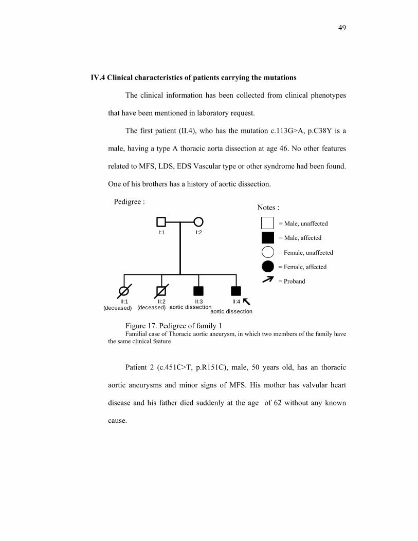
IV.4 Clinical characteristics of patients carrying the mutations
The clinical information has been collected from clinical phenotypes
that have been mentioned in laboratory request.
The first patient (II.4), who has the mutation c.113G>A, p.C38Y is a
male, having a type A thoracic aorta dissection at age 46. No other features
related to MFS, LDS, EDS Vascular type or other syndrome had been found.
One of his brothers has a history of aortic dissection.
Pedigree :
Figure 17. Pedigree of family 1 Familial case of Thoracic aortic aneurysm, in which two members of the family have
the same clinical feature
Patient 2 (c.451C>T, p.R151C), male, 50 years old, has an thoracic
aortic aneurysms and minor signs of MFS. His mother has valvular heart
disease and his father died suddenly at the age of 62 without any known
cause.
Notes :
= Male, unaffected
= Male, affected
= Female, unaffected
= Female, affected
= Proband
II:4
I:1 I:2
II:3II:1 II:2
aortic dissectionaortic dissection(deceased)(deceased)
50
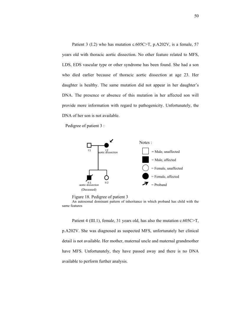
Patient 3 (I.2) who has mutation c.605C>T, p.A202V, is a female, 57
years old with thoracic aortic dissection. No other feature related to MFS,
LDS, EDS vascular type or other syndrome has been found. She had a son
who died earlier because of thoracic aortic dissection at age 23. Her
daughter is healthy. The same mutation did not appear in her daughter’s
DNA. The presence or absence of this mutation in her affected son will
provide more information with regard to pathogenicity. Unfortunately, the
DNA of her son is not available.
Pedigree of patient 3 :
Figure 18. Pedigree of patient 3 An autosomal dominant pattern of inheritance in which proband has child with the
same features
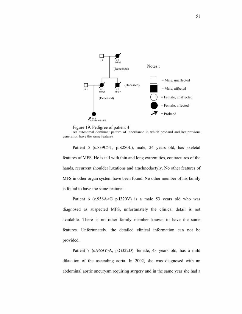
Patient 4 (III.1), female, 31 years old, has also the mutation c.605C>T,
p.A202V. She was diagnosed as suspected MFS, unfortunately her clinical
detail is not available. Her mother, maternal uncle and maternal grandmother
have MFS. Unfortunately, they have passed away and there is no DNA
available to perform further analysis.
I:2I:1
II:1 II:2
aortic dissection
aortic dissection
(Deceased)
Notes :
= Male, unaffected
= Male, affected
= Female, unaffected
= Female, affected
= Proband
51
Figure 19. Pedigree of patient 4 An autosomal dominant pattern of inheritance in which proband and her previous
generation have the same features
Patient 5 (c.839C>T, p.S280L), male, 24 years old, has skeletal
features of MFS. He is tall with thin and long extremities, contractures of the
hands, recurrent shoulder luxations and arachnodactyly. No other features of
MFS in other organ system have been found. No other member of his family
is found to have the same features.
Patient 6 (c.958A>G p.I320V) is a male 53 years old who was
diagnosed as suspected MFS, unfortunately the clinical detail is not
available. There is no other family member known to have the same
features. Unfortunately, the detailed clinical information can not be
provided.
Patient 7 (c.965G>A, p.G322D), female, 43 years old, has a mild
dilatation of the ascending aorta. In 2002, she was diagnosed with an
abdominal aortic aneurysm requiring surgery and in the same year she had a
III:1
II:1 II:2
I:1 I:2
II:3
suspected MFS
MFS? MFS?
MFS?
Notes :
= Male, unaffected
= Male, affected
= Female, unaffected
= Female, affected
= Proband
(Deceased)
(Deceased)
(Deceased)
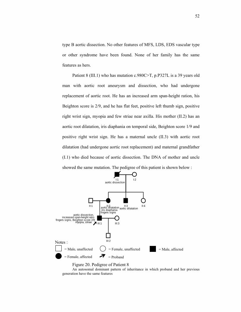
52
Notes :
type B aortic dissection. No other features of MFS, LDS, EDS vascular type
or other syndrome have been found. None of her family has the same
features as hers.
Patient 8 (III.1) who has mutation c.980C>T, p.P327L is a 39 years old
man with aortic root aneurysm and dissection, who had undergone
replacement of aortic root. He has an increased arm span-height ration, his
Beighton score is 2/9, and he has flat feet, positive left thumb sign, positive
right wrist sign, myopia and few striae near axilla. His mother (II.2) has an
aortic root dilatation, iris diaphania on temporal side, Beighton score 1/9 and
positive right wrist sign. He has a maternal uncle (II.3) with aortic root
dilatation (had undergone aortic root replacement) and maternal grandfather
(I.1) who died because of aortic dissection. The DNA of mother and uncle
showed the same mutation. The pedigree of this patient is shown below :
Figure 20. Pedigree of Patient 8 An autosomal dominant pattern of inheritance in which proband and her previous
generation have the same features
I:1 I:2
II:2 II:3 II:4II:1
III:1 III:3
III:2
aortic dissection, increased span-height ratio
fingers signs, Beighton score 2/9myopia, striae
aortic dilatationiris diaphania aortic dilatationfingers signs
aortic dissection
= Male, unaffected = Male, affected = Female, unaffected
= Female, affected = Proband

53
Patient 9 (II.1) who has mutation c.1282T>G, p.Y428D is male, 45
years old diagnosed as having thoracic aortic aneurysm and dissection at the
age of 35 years. His mother has a descending aortic aneurysm. No other
features of MFS, LDS, EDS vascular type or other syndromes have been
found.
Figure 21. Pedigree of patient 9 An autosomal dominant pattern of inheritance in which proband and have the same clinical feature
Patient 10, (c.1460G>A, p.R487Q), female, 17 years old (IV.2), was
diagnosed as having an aortic aneurysm and hypermobility of the joints at
10 years old. Her brother (IV.1) carries the same mutation and also having
aortic aneurysms. No other features of MFS were apparent in these two
patients. The pedigree is depicted in figure 7. I.2 died of aortic dissection at
the age of 36 years, II-2 at the age of 50 years, III-2 at the age of 21 years
II:1
I:1 I:2
II:2
III:1III:2
descenden aortic aneurysm
aortic aneurysm & dissection
Notes :
= Male, unaffected
= Male, affected
= Female, unaffected
= Female, affected
= Proband
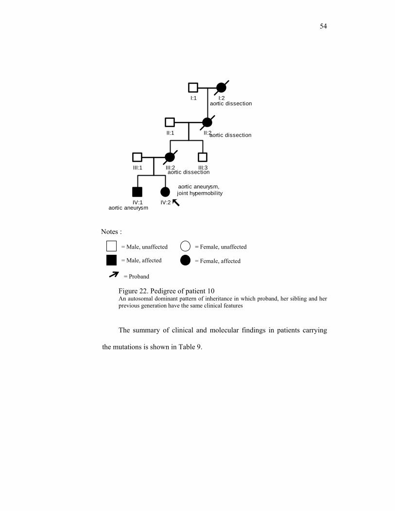
54
Figure 10. Pedigree of patient 10
Figure 22. Pedigree of patient 10 An autosomal dominant pattern of inheritance in which proband, her sibling and her previous generation have the same clinical features
The summary of clinical and molecular findings in patients carrying
the mutations is shown in Table 9.
I:1 I:2
II:2II:1
III:2 III:3III:1
IV:1 IV:2aortic aneurysm
aortic aneurysm,joint hypermobility
aortic dissection
aortic dissection
aortic dissection
Notes :
= Male, unaffected
= Male, affected
= Female, unaffected
= Female, affected
= Proband
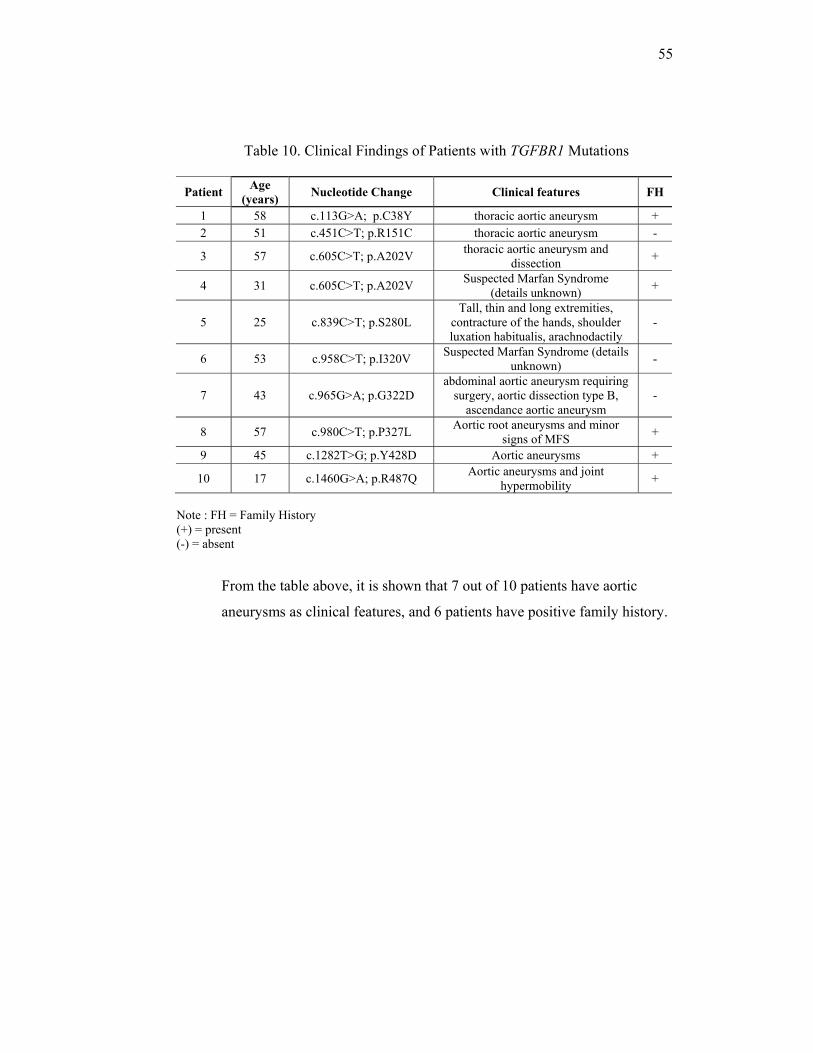
55
Table 10. Clinical Findings of Patients with TGFBR1 Mutations
Patient Age (years) Nucleotide Change Clinical features FH
1 58 c.113G>A; p.C38Y thoracic aortic aneurysm + 2 51 c.451C>T; p.R151C thoracic aortic aneurysm -
3 57 c.605C>T; p.A202V thoracic aortic aneurysm and dissection +
4 31 c.605C>T; p.A202V Suspected Marfan Syndrome (details unknown) +
5 25 c.839C>T; p.S280L Tall, thin and long extremities,
contracture of the hands, shoulder luxation habitualis, arachnodactily
-
6 53 c.958C>T; p.I320V Suspected Marfan Syndrome (details unknown) -
7 43 c.965G>A; p.G322D abdominal aortic aneurysm requiring
surgery, aortic dissection type B, ascendance aortic aneurysm
-
8 57 c.980C>T; p.P327L Aortic root aneurysms and minor signs of MFS +
9 45 c.1282T>G; p.Y428D Aortic aneurysms +
10 17 c.1460G>A; p.R487Q Aortic aneurysms and joint hypermobility +
Note : FH = Family History (+) = present (-) = absent
From the table above, it is shown that 7 out of 10 patients have aortic
aneurysms as clinical features, and 6 patients have positive family history.
56
Chapter V
DISCUSSION
Most of the patients included in this study were those with suspected MFS
with or without the presence of aortic aneurysm and/or dissection (78/194), aortic
aneurysm and/or dissection (60/194), and familial cases of aortic aneurysm and/or
dissection (42/194). The mutations found in this study is 10 mutations in 194
samples (10/194). Previously published results by Singh et al (2006) and Loeys et
al (2006) on MFS patients without FBN1 mutations, as reviewed by Mizuguchi T
and Matsumoto N, yield 2/41 and 1/22, respectively.34 The study by Matyas et al
(2006) has found 4.0% (3/70) TGFBR1 mutations on MFS-related patients
without FBN1 involvement.11 Furthermore, combining the findings of TGFBR1
mutations in LDS and Thoracic Aortic Aneurysms and Dissections (TAAD), it is
very likely that TGFBR1 mutations do play role in the pathogenesis of MFS and
related disorders through TGFβ signaling, although the frequency is not
significant. Mutation analysis on TGFBR1 should be considered in MFS and its
related disorders, without the presence of mutations in FBN1 and TGFBR2.
All the mutations that have been found in this study are novel, except one
mutation in exon 9 p.R487Q, so there is no previous publication about the
pathogenicity of these mutations. The pathogenicity of mutations were analyzed
based on the changes in amino acid, the amino acid conservation across the
species, the protein domain where the mutation was occurred, in some patients we
looked for the presence or absence of the mutation in affected or unaffected
57
family members respectively, and use internet-based prediction tools software
PolyPhen and SIFT. The mutation p.C38Y is considered pathogenic from the
multiple alignment analysis and PolyPhen analysis. The result from PolyPhen
analysis predicted that the changes will disturb the formation of disulfide bond of
the protein, thus will disturb the structure of the protein. The mutation p.R151C is
predicted to be benign and tolerated by Polyphen and SIFT analysis, respectively.
However, considering big changes in amino acid type and the domain
conservation across multiple species, this mutation is considered as pathogenic.
The mutation c.605C>T, p.A202V has been predicted to affect protein function
based on SIFT analysis, but predicted as benign on PolyPhen. It occurred at a
highly conserved domain. However, this mutation is unlikely to be pathogenic
because the change in amino acid type is not significant. The mutation p.S280L is
considered pathogenic because the change from serine to leucine is a significant
change. Serine and threonine residues can be autophosphorylated, but not leucine.
The mutation p.I320V occurred at highly conserved domain, predicted as possibly
damaging by PolyPhen analysis and “tolerated” by SIFT analysis. However, this
mutation is unlikely to be pathogenic because the change in amino acid type is
not significant. The mutation p.G322D is predicted to be benign and tolerated on
PolyPhen and SIFT analysis. But it is considered pathogenic, because of the
significant change in amino acid, the conservation in 11 different species and the
occurrence in the protein kinase domain. In patient with mutation p.P327L, the
DNA of affected mother and affected uncle showed the same mutation. This
mutation is predicted to be probably damaging and affects protein function by
58
both PolyPhen and SIFT analysis. The location of this mutation in protein kinase
domain and the high conservation across 11 species, make this mutation strongly
suggested as pathogenic. The mutation p.Y428D is predicted to be probably
damaging and affects protein function by PolyPhen and SIFT analysis,
respectively. With regard to a big change in amino acid, this mutation is
considered pathogenic.
Mutation p.R487Q has been found previously to be pathogenic in other
studies.10,11,35 Akutsu et al (2007) found this mutation in patient with acute aortic
dissection, mesenteric artery aneurysm and bilateral pneumothorax, without other
features of MFS or LDS.34 Matyas et al (2006) found this mutation in patient with
thoracic aortic aneurysm and dissection, also without any features of MFS and
LDS.11 Loeys et al (2006) described this mutation in patients with thoracic aortic
aneurysm and thoracic aortic dissection, without other features of MFS or LDS.10
From those three previous publications, it seems that a mutation in this location
causes aortic aneurysm and dissection, and is not likely to cause skeletal or eye
abnormality.
Seven out of nine different mutations in this study occurred at highly
conserved kinase domains, more specifically, the serine-threonine kinase domain.
This domain is responsible for the formation of kinase, an enzyme that plays a
role in cellular processes, including division, proliferation, apoptosis and
differentiation.35 Most of the mutations in TGFBR1 that have been published are
located in this domain.10,11,13,20 Thus, it is strongly suggested that this domain has
59
a very crucial role in the formation of TGFβR1 and mutations in this domain are
pathogenic.
A polymorphism in exon 1 (c.70_78delGCGGCGGCG), the 6Ala allele,
was predicted to act as low penetrance allele of the clinical features in Marfan
Syndrome.37 This 6Ala allele has been previously associated with a higher risk of
colorectal cancer, breast cancer and ovarian cancer. 6Ala/6Ala Homozygosity
even leads to higher risk than 6Ala/9Ala heterozygosity.38 Whether it acts in the
same way in MFS and related disorders, however, needs a broader analysis which
includes a large number of controls.
Among 194 patients with MFS and related disorders, we found 10 patients
carrying TGFBR1 mutations. Based on the data available, none of these patients
was diagnosed clinically as MFS fulfilling the Ghent criteria, nor had features of
LDS or other syndromes. These diseases have common nature, that the features
might be shown to be age-dependent, thus a clinical follow up should be provided
to confirm present diagnosis. In this study, all patients are more than 17 years old.
Since they already exceed pubertal ages, the chance for developing new feature is
not likely. But the existing feature still should be monitored for developing worse.
From 10 cases with TGFBR1 mutations (7 pathogenics, 3 non
pathogenics), seven out of ten patients with TGFBR1 mutation have aortic
aneurysm, with 3 of them also have minor features of MFS. Seven of them are
familial cases. The exact phenotype due to TGFBR1 mutations cannot be clearly
concluded, since these patients have features ranging from isolated aortic
aneurysm to skeletal abnormalities. Singh et al found TGFBR1 mutations in
60
patients with typical Marfan Syndrome.19 Loeys et al, Akutsu et al, Drera et al
found mutations in patients with features of Loeys-Dietz syndrome.21,35,39 Ades et
al reported mutations in Furlong Syndrome.12 Matyas et al found it in even larger
variety of clinical features variation: in TAAD, LDS and typical MFS patients.11
Thus, it is suggested that mutations in TGFBR1 have variable clinical outcomes,
indeed. The likeliness of patients with TGFBR1 mutation having aortic aneurysms
might be one sign that lead us to do TGFBR1 mutation analysis in MFS and
related disorders patient. It has been recognized also, that mutation in TGFBR
genes rarely found in classic type of MFS. In this study we found none of patient
with classic MFS has mutation in TGFBR.
Marfan Syndrome, Loeys-Dietz Syndrome, Familial Thoracic Aortic
Aneurysms and Dissections and other Marfan-related disorders are inherited in
autosomal dominant manner. An individual whose parent is carrying a
heterozygous mutation of the gene causing this disorder will have 50% chance to
develop the disorder. In this study, 6 familial cases with TGFBR1 mutations have
been found. In these families, genetic counseling should be provided to inform
them about the risk and how to deal with the disorders in the future. The nature of
MFS and aortic aneurysms is that the clinical presentations develops with age.
Patients must be warned and counseled that once they are diagnosed, they need to
be monitored for aortic widening, skeletal growth, etc. The medicinal treatment
needs to be taken a life long, and surgical treatment may be needed.
Furthermore, mutations in TGFBR genes are related with more severe
vascular manifestation with probably a shortened life expectancy.7 In EDS
61
vascular type, there’s a shortened survival, with the mean age is 48 years. The
presence of tissue fragility make it difficult to do aorta repair, and increase the risk
of visceral rupture.40 Patients affected by Loeys-Dietz Syndrome have high risk of
aortic aneurysm and dissection. But the repairing surgery is usually less
complication and more successful in LDS compared to EDS vascular type.10 In
this case, a careful recognition on clinical presentation and molecular examination
play an important role.
In the rest of the 184 patients, the causative gene responsible for their
disorders has been left unknown. Further research on genes which play role in the
TGF-β signaling pathway, such as LTBP4, or TGFB itself9 will be needed. In
patients having aortic aneurysms, other candidate genes which play role in
maintaining the aortic structure, such as COL3A1, ACTA2 and MYH1141,42 may
also be involved.
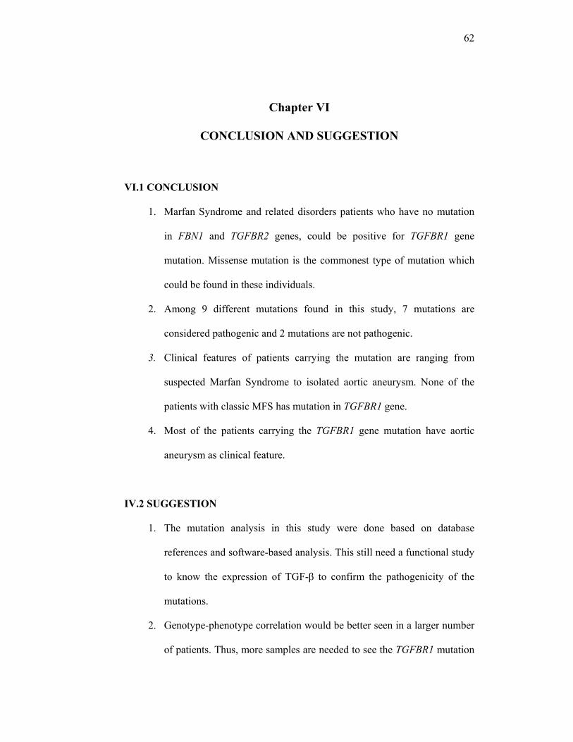
62
Chapter VI
CONCLUSION AND SUGGESTION
VI.1 CONCLUSION
1. Marfan Syndrome and related disorders patients who have no mutation
in FBN1 and TGFBR2 genes, could be positive for TGFBR1 gene
mutation. Missense mutation is the commonest type of mutation which
could be found in these individuals.
2. Among 9 different mutations found in this study, 7 mutations are
considered pathogenic and 2 mutations are not pathogenic.
3. Clinical features of patients carrying the mutation are ranging from
suspected Marfan Syndrome to isolated aortic aneurysm. None of the
patients with classic MFS has mutation in TGFBR1 gene.
4. Most of the patients carrying the TGFBR1 gene mutation have aortic
aneurysm as clinical feature.
IV.2 SUGGESTION
1. The mutation analysis in this study were done based on database
references and software-based analysis. This still need a functional study
to know the expression of TGF-β to confirm the pathogenicity of the
mutations.
2. Genotype-phenotype correlation would be better seen in a larger number
of patients. Thus, more samples are needed to see the TGFBR1 mutation
63
frequency in higher population. The stricter inclusion criteria will be
expected to have better conclusion in this matter, too.
3. Better study design in which we can follow the disease progress in MFS
and related disorders patients should be used to get better understanding
on the involvement and the impact of TGFBR1 mutations in MFS and
related disorders pathogenesis and severity.
4. This field of research has large potential to be explored in Indonesian
population. The DNA samples from Indonesian patients is needed to
perform the research. Centers which provide DNA sequencing services
should be available.
5. When the cost of DNA sequencing becomes a major problem, mutation
screening methods, such as MLPA (Multiplex Ligation Dependent Probe
Amplification) may be considered. By mutation screening methods, the
requirement for DNA sequencing can be significantly reduced.
6. Mutation analysis in other genes should be done on the rest of patients
whose cause of the disorders are still unknown.
64
Chapter VII
SUMMARY
Marfan Syndrome (MFS), a common autosomal dominant inherited disorder
of fibrous connective tissue, mostly affects three organ systems : skeletal, ocular
and cardiovascular system. Cardiovascular involvements, the aortic aneurysms
leading to aortic dissection or rupture, is the most life-threatening.
Diagnosis of MFS can be established by the Ghent criteria. However, the
interpretation of these criteria is not always easy, due to the presence of many
disorders which are clinically similar to MFS. Those disorders, termed as related
disorders of MFS include Loeys-Dietz syndrome, Sphrintzen-Goldberg
Syndrome, Familial Aortic Aneurysm, Bicuspid Aortic Valve with Aortic
Dilatation, Familial Ectopia Lentis, MASS phenotype, Marfan Body Type, Mitral
Valve Prolapse Syndrome, Congenital Contractural Arachnodactily (Beals
syndrome), Stickler syndrome and Ehlers-Danlos syndrome.
Previously, the pathogenesis of MFS was explained based on the concept of
fibrillin-rich micro fibrils as purely architectural elements in the extra cellular
matrix. Mutations in the fibrillin-1 gene (FBN1 gene), known to cause MFS,
however, the mutations have not always been found in MFS patients.
Recent findings on the pathogenesis of MFS demonstrate changes in growth
factor signaling and other changes in matrix-cell interactions. Mouse models of
MFS with FBN1 mutation which have lung emphysema as phenotypic
manifestation, showed increased TGFβ signalling. The involvement of TGFβ-
65
receptor gene mutation in MFS has been shown in a Japanese patient with MFS
who had a balanced chromosomal translocation involving chromosome 3p24. This
locus had been found to show genetic linkage with MFS in a large French
pedigree. The breakpoint in the Japanese patient disrupted the TGFBR2 gene.
The proteins fibrillin-1, TGFBR1 and TGFBR2 take part in transforming
growth factors β (TGFβ) signaling, thus mutations in one of these gene could
cause similar phenotypes. Mutation analysis on FBN1 and TGFBR2 genes in MFS
and related disorders have been well established, and is important to distinguish
those Marfan spectrum disorders from one and another. However, since there are
still many cases without any mutation in either FBN1 or TGFBR2, mutation
analysis on other candidate gene is needed to be performed. Mutation analysis on
TGFBR1 gene as one of candidate gene, which include the recognition of
mutation and its kind, the prediction on pathogenicity, the distribution of
phenotypes on genotypes and the recognition of clinical sign which may lead to
this gene, are need to be done.
The TGFBR1 gene is also known as activin A receptor like kinase, or serine
/ threonine-protein kinase receptor R4 gene. The DNA size is approximately 45kb
long, the mRNA size is 2308bp, contains of 9 exons and is located on
chromosome 9q22.33. The protein domains of TGFBR1 consist of extra cellular
domain, transmembrane domain, cytoplasmic domain, glycine-serine rich domain,
and serine-threonine kinase domain. These domains are highly conserved across
species.
66
This research is in the field of medical genetics, held in the DNA Diagnostic
Laboratory of Vrije Universiteit Medisch Centrum (VUmc), Amsterdam, The
Netherlands. This is a descriptive study. The population of this research is the
DNA samples of patients with Marfan Syndrome and related disorders which have
been reffered to DNA Diagnostic Laboratory of VUmc Hospital Amsterdam, The
Netherlands from the year 1998-2008. The DNA samples were donation with
permission from Gerard Pals, PhD as the principal investigator of Connective
Tissue Disorders research in the DNA Diagnostic Laboratory of VUmc Hospital
Amsterdam, The Netherlands. All of the samples used in this research are part of
Connective Tissue Disorders research project, and have been consent to be
included in research.
One hundred and ninety four DNA of unrelated patients with MFS,
suspected MFS, or related disorders, have been included. The inclusion criteria
were having at least one major criterion of MFS and found to be negative for
FBN1 and TGFBR2 mutations on previous examination. The samples were
excluded if the amount of the DNA were not enough for further analysis. The
phenotypic characteristics of the patients were then traced from their laboratory
request form.
PCR was done to amplify the whole 9 exons of TGFBR1. The PCR products
were then confirmed by gel electrophoresis, and underwent a pre-sequencing
preparation before go to an automated sequencing machine. The results of the
DNA sequencing were then analyzed for the presence of variants. When a variant
has been found, the database of mutations and polymorphisms would be used to
67
confirm whether the variant is a mutation or polymorphism. When it was not in
the database, then the following things would be considered to decide the
pathogenicity : the amino acid changes, domain localization, conservation across
species using multiple sequence alignment, and the prediction results from
internet-based software : PolyPhen and SIFTblink.
The patients were grouped into several diagnoses based on clinical findings
and matched with Ghent Criteria. A diagnosis of MFS was based on Ghent
Criteria. Incomplete Ghent Criteria, or having at least one major criterion in an
organ system with minor criterion of another organ, or more than one minor
criterion, would be considered as Suspected MFS. The patients with only specific
clinical features (such as only has aortic aneurysm, ectopia lentis, dural ectasia or
joint hypermobility) would be grouped as the clinical findings, recognized as
Marfan Syndrome, Suspected MFS, Aortic Aneurysms and/ Dissections, Familial
Aortic Aneurysms and/ Dissections, Ectopia Lentis, Dural Ectasia, Joint
Hypermobility. There are 10 MFS, 78 Suspected MFS, 60 Aortic aneurysms and/
Dissections, 42 Familial aortic aneurysms and dissections, 2 Ectopia Lentis, 1
Dural ectasia and 1 joint hypermobility patients.
On sequencing all 9 exons of TGFBR1, a total of 9 mutations, 7 different
polymorphisms and 3 unclassified variants in TGFBR1 were found. The mutations
were found in 10 patients. The 9 mutations, occured in 7 different exons.
The first mutation c.113G>A; p.C38Y is located in exon 2 of TGFBR1 gene,
at the position 113 of cDNA, in which guanine is replaced by adenine, resulted in
the change of amino acid 38 from cysteine (a polar-neutral amino acid) to tyrosine
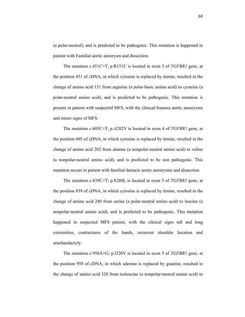
68
(a polar-neutral), and is predicted to be pathogenic. This mutation is happened in
patient with Familial aortic aneurysm and dissection.
The mutation c.451C>T; p.R151C is located in exon 3 of TGFBR1 gene, at
the position 451 of cDNA, in which cytosine is replaced by timine, resulted in the
change of amino acid 151 from arginine (a polar-basic amino acid) to cysteine (a
polar-neutral amino acid), and is predicted to be pathogenic. This mutation is
present in patient with suspected MFS, with the clinical features aortic aneurysms
and minor signs of MFS.
The mutation c.605C>T; p.A202V is located in exon 4 of TGFBR1 gene, at
the position 605 of cDNA, in which cytosine is replaced by timine, resulted in the
change of amino acid 202 from alanine (a nonpolar-neutral amino acid) to valine
(a nonpolar-neutral amino acid), and is predicted to be non pathogenic. This
mutation occurs in patient with familial thoracic aortic aneurysms and dissection.
The mutation c.839C>T; p.S280L is located in exon 5 of TGFBR1 gene, at
the position 839 of cDNA, in which cytosine is replaced by timine, resulted in the
change of amino acid 280 from serine (a polar-neutral amino acid) to leucine (a
nonpolar-neutral amino acid), and is predicted to be pathogenic. This mutation
happened in suspected MFS patient, with the clinical signs tall and long
extremities, contractures of the hands, recurrent shoulder luxation and
arachnodactyly.
The mutation c.958A>G; p.I320V is located in exon 5 of TGFBR1 gene, at
the position 958 of cDNA, in which adenine is replaced by guanine, resulted in
the change of amino acid 320 from isoleucine (a nonpolar-neutral amino acid) to
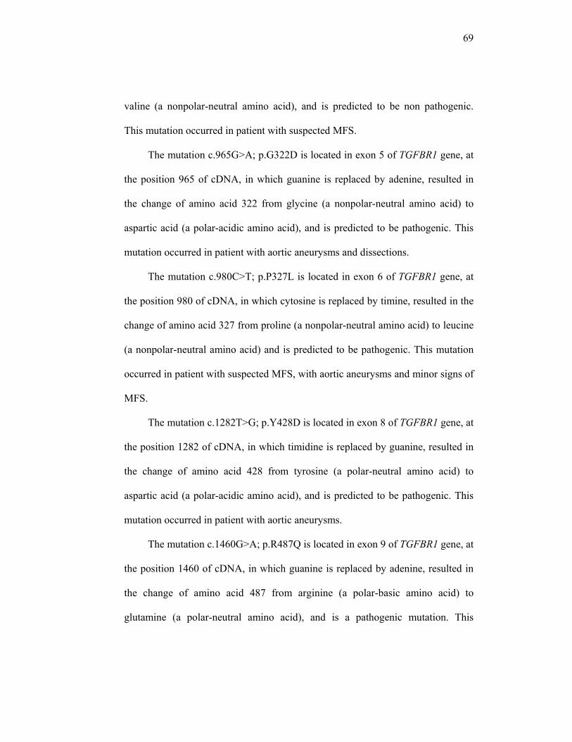
69
valine (a nonpolar-neutral amino acid), and is predicted to be non pathogenic.
This mutation occurred in patient with suspected MFS.
The mutation c.965G>A; p.G322D is located in exon 5 of TGFBR1 gene, at
the position 965 of cDNA, in which guanine is replaced by adenine, resulted in
the change of amino acid 322 from glycine (a nonpolar-neutral amino acid) to
aspartic acid (a polar-acidic amino acid), and is predicted to be pathogenic. This
mutation occurred in patient with aortic aneurysms and dissections.
The mutation c.980C>T; p.P327L is located in exon 6 of TGFBR1 gene, at
the position 980 of cDNA, in which cytosine is replaced by timine, resulted in the
change of amino acid 327 from proline (a nonpolar-neutral amino acid) to leucine
(a nonpolar-neutral amino acid) and is predicted to be pathogenic. This mutation
occurred in patient with suspected MFS, with aortic aneurysms and minor signs of
MFS.
The mutation c.1282T>G; p.Y428D is located in exon 8 of TGFBR1 gene, at
the position 1282 of cDNA, in which timidine is replaced by guanine, resulted in
the change of amino acid 428 from tyrosine (a polar-neutral amino acid) to
aspartic acid (a polar-acidic amino acid), and is predicted to be pathogenic. This
mutation occurred in patient with aortic aneurysms.
The mutation c.1460G>A; p.R487Q is located in exon 9 of TGFBR1 gene, at
the position 1460 of cDNA, in which guanine is replaced by adenine, resulted in
the change of amino acid 487 from arginine (a polar-basic amino acid) to
glutamine (a polar-neutral amino acid), and is a pathogenic mutation. This

70
mutation occurred in patient with aortic aneurysms and dissection with joint
hypermobility.

71
REFERENCES
1. OMIM (Online Mendelian Inheritance in Man). Marfan Syndrome #154700.
Available at URL : http://www.ncbi.nlm.nih.gov/entrez/dispomim.cgi?id=154700
2. Colloud-Beroud G, Boileau C. Marfan syndrome in the third millenium. Eur J Hum Genet. 2002; 10 : 673-681
3. Millewicz DM, Dietz HC, Miller DC. Treatment of aortic disease in patients with Marfan Syndrome. Circulation. 2005 ; 111 : e150-e157
4. Robinson PN, Arteaga-Solis E, Baldock C, Collod-Beroud G, Booms P, De Paepe A, et al. The molecular genetics of marfan syndrome and related disorders. Journal of Medical Genetics 2006; 43 : 769-787
5. De Paepe A, Devereux RB, Dietz HC, Hennekam RCM, Pyeritz RE. Revised diagnostic criteria of marfan syndrome. American Journal of Medical Genetics. 1996 ; 62 : 417-476
6. Neptune ER, Frischmeyer PA, Arking DE, Myers L, Bunton T, Gayraud B, et al. Dysregulation of TGFβ activation contributes to pathogenesis in marfan syndrome. Nature Genetics. 2003 (33); 407-411
7. Mizuguchi T, Collod-Beroud G, Akiyama T, Abifadel M, Harada N, Morisaki T, et al. Heterozygous TGFBR1 mutations in marfan syndrome. Nature Genetics Advance Online Publication. 2004. doi:10.1038/ng1392
8. Habashi JP, Judge DP, Holm TM, Cohn RD, Loeys BL, Cooper TK, et al. Losartan, an AT1 antagonist, prevents aortic aneurysm in a mouse model of marfan syndrome. Science. 2006; 312 : 117
9. Ten Dijke P, Arthur HM. Extracellular control of TGFβ signalling in vascular development and diseases. Nature Reviews on Molecular Cell Biology. 2007 ; 7 : 857-869
10. Loeys BL, Schwarze U, Holm T, Callewaert BL, Thomas GH, Pannu H, et al. Aneurysm syndromes caused by mutations in the TGF-beta receptor. N Engl J Med. 2006 Aug 24;355(8):788-98.
11. Matyas G, Arnold E, Carrel T, Baumgartner D, Boileau C, Berger W, Steinmann B. Identification and in silico analysis of novel TGFβR1 and TGFβR2 in MFS-related disorders. Hum Mutat. 2006 ; 27(8) : 760-769

72
12. Ades LC, Sullivan K, Biggin A, Haan EA, Brett M, Holman KJ, et al. FBN1, TGFBR1, and the Marfan-craniosynostosis/mental retardation disorders revisited. Am J Med Genet A. 2006 May 15;140(10):1047-58.
13. UMD (Universal Mutation Database) for TGFBR1 mutations. Available at URL: http://www.umd.be
14. Dietz, HC. Gene Review : Marfan syndrome, 2005. Available at http://www.genetesthttp://www.genetests.org/servlet/access?db=geneclinics&site=gt&id=8888891&key=eK2Jaf8ktIuY&gry=&fcn=y&fw=fcPS&filename=/profiles/marfan/index.html
15. Dean, JCS. Marfan Syndrome : clinical diagnosis and management. Eur J Hum Genet. 2007; 15:724-733
16. GenAtlas : TGFBR1. Available at URL : http://genatlas.medecine.univ-paris5.fr/fiche.php?symbol=TGFBR1
17. OMIM (Online Mendelian Inheritance in Man). Transforming growth factor-beta receptor, type I ; TGFBR1*190181. Available at URL : http://www.ncbi.nlm.nih.gov/entrez/dispomim.cgi?id=190181
18. Ace view : a comprehensive cDNA-supported genes and transcript annotation, Genome Biology 2006, 7(Suppl 1):S12
19. Singh KK, Rommel K, Mishra A, Karck M, Haverich A, Schmidtke J, Arslan-Kirchner M. TGFBR1 and TGFBR2 mutations in patients with features of Marfan syndrome and Loeys-Dietz syndrome. Hum Mutat. 2006 Aug;27(8):770-7.
20. Loeys Bl, Chen J, Neptune ER, Judge DP, Podowski M, Holm T, et al. A syndrome of altered cardiovascular, craniofacial, neurocognitive and skeletal development caused by mutations in TGFBR1 or TGFBR2 (Letters). Nature Genetic. 2005; 37(3), 275-281
21. Kosaki K, Takahashi D, Udaka T, Kosaki R, Matsumoto T, Ibe S, et al. Molecular pathology of Sphrintzen-Goldberg syndrome. Am J Med Genet A. 2006; 140:104-10
22. Bell J, Bodmer D, Sistermans E, Ramsden SC. Practice guidelines for the interpretation and reporting of unclassified variants (Uvs) in clinical molecular genetics. UK Clinical Molecular Genetics Society (CMGS) and The Dutch
73
Society of Clinical Genetic Laboratory Specialists (Vereniging Klinisch Genetische Laboratoriumspecialisten; VKGL). 2007.
23. Strachan T, Read AP. Human molecular genetics 3. Garland Publishing, London and New York. 2004; 317-321.
24. Pauline C Ng, Steven Henikoff. Predicting the effects of amino acid substitutions on protein function. Annu Rev. Genomics Hum.Genet.2006.7:61-80
25. PolyPhen : prediction of functional effect of human nsSNPs. Available at URL : http://coot.embl.de/PolyPhen/
26. Polyphen help. Available at URL : http://genetics.bwh.harvard.edu/pph/pph_help.html
27. Sorting Intolerant From Tolerant (SIFT) Available at URL : http://blocks.fhcrc.org/sift/SIFT_BLink_submit.html
28. Madiyono B. Perkiraan besar sampel. In Dasar-Dasar Metodologi Klinis. Editor: Sastroasmoro,et.al. Second Edition. Sagung Seto, Jakarta: p:270
29. Gene sequence information for ENSG00000106799. Available at URL : http://www.ensembl.org/Homo_sapiens/geneseqview?db=core;gene=ENSG00000106799
30. Ensembl transcript report. Available at URL : http://www.ensembl.org/Homo_sapiens/transview?db=core;transcript=ENST00000374994
31. UniprotKB/Swiss-Prot P36897 (TGF-beta R_Human). Available at URL : http://www.uniprot.org/uniprot/P36897#section_features
32. Human Splicing Finder. Available at URL : http://www.umd.be/HSF/ 33. Akutsu K, Morisaki H, Takeshita S, Sakamoto S, Tamori Y, Yoshimuta T, et
al. Phenotypic heterogeneity of Marfan-like connective tissue disorders associated with mutations in the transforming growth factor-beta receptor genes. Circ J. 2007 Aug;71(8):1305-9.
34. Sakai H, Visser R, Ikegawa S, Ito E, Numabe H, Watanabe Y, et al. Comprehensive genetic analysis of relevant four genes in 49 patients with Marfan syndrome or Marfan-related phenotypes. Am J Med Genet A. 2006 Aug 15;140(16):1719-25.

74
35. TGFBR1 HomoloGene. Available at URL : http://www.ncbi.nlm.nih.gov/sites/entrez?cmd=Retrieve&db=homologene&dopt=MultipleAlignment&list_uids=3177
36. Mizuguchi T, Matsumoto N. Recent progress in genetics of Marfan Syndrome and Marfan-associated disorders. J Hum Genet (2007) 52:1-12
37. Manning G, Plowman GD, Hunter T, Sudarsanam S. Evolution of protein kinase signaling from yeast to man (Review). TRENDS in Biochemical Sciences (2002) 27(10) : 514-520
38. Lucarini L, et al. May TGFBR1 act also as low penetrance allele in Marfan Syndrome? Int J Cardiol (2007), doi:10.1016/j.ijcard.2007.07.048
39. De la Chapelle, A. Genetic predisposition to colorectal cancer. Nature Reviews : Cancer (2004) 4:769-780
40. Drera B, Tadini G, Barlati S, Colombi M. Identification of a novel TGFBR1 mutation in a Loeys-Dietz syndrome type II patient with vascular Ehlers-Danlos syndrome phenotype. Clin Genet. 2007 Dec 6;. [Epub ahead of print]
41. Child AH, Neumann L, Robinson PN. Diagnosis and treatment of Marfan Syndrome—a summary, in : Marfan Syndrome : A Primer for Clinicians and Scientists. Kluwer Academics / Plenum Publisher. New York : 2004; 13-23
42. Pepin MMS, Schwarze U, Superti-Furga A, Byers PH. Clinical and genetic features of Ehlers-Danlos Type IV, the vascular type. N Engl J Med. 2000 ; 342(10):673-680
43. Patel HJ, Deeb GM. Ascending and arch aorta : pathology, natural history and treatment. Circulation. 2008;118:188-195
44. Millewicz DM, Guo DC, Tran-Fadulu V, Lafont AL, Papke CL, Inamoto S, Pannu H. Genetic basis of thoracic aortic aneurysms and dissections : focus on smooth muscle contractile dysfunction. Annu. Rev. Genomic Hum. Genet. 2008; 9:283-30
45. Krivokapich J, Child JS, Dadourian BJ, Perloff JK. Reassessment of echocardiographic criteria for diagnosis of mitral valve prolapse. Am J Cardiol. 1988; 61:131-135
46. Sponseller P, Shindle M. Orthopedic Problems in marfan Syndrome., in Marfan Syndrome : A Primer for Clinicians and Scientists. Kluwer Academics / Plenum Publisher. New York : 2004; 26-27

75
47. Von Kodolitsch Y, Rybczynski M. Cardiovascular aspect in marfan syndrome : a systematic review. In : Marfan Syndrome : A Primer for Clinicians and Scientists. Medical Intelligent Unit, Plenum Publisher. 2004;45-69
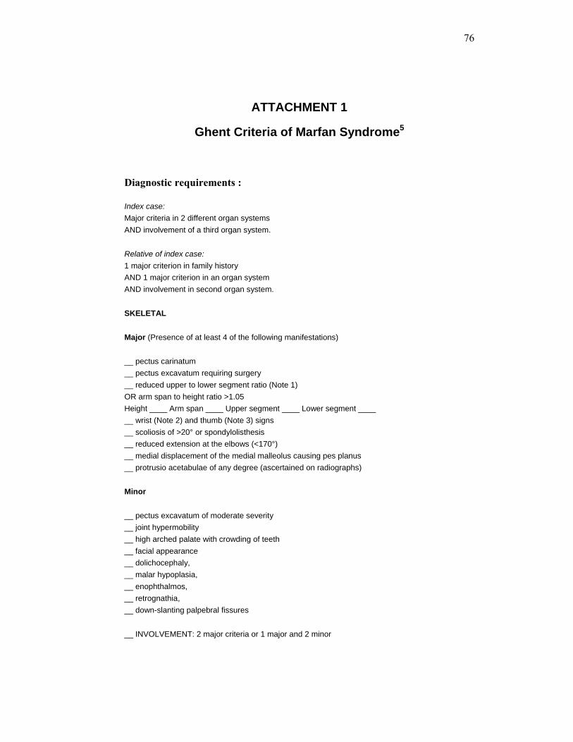
76
ATTACHMENT 1
Ghent Criteria of Marfan Syndrome5
Diagnostic requirements :
Index case: Major criteria in 2 different organ systems AND involvement of a third organ system. Relative of index case: 1 major criterion in family history AND 1 major criterion in an organ system AND involvement in second organ system. SKELETAL Major (Presence of at least 4 of the following manifestations) __ pectus carinatum __ pectus excavatum requiring surgery __ reduced upper to lower segment ratio (Note 1) OR arm span to height ratio >1.05 Height ____ Arm span ____ Upper segment ____ Lower segment ____ __ wrist (Note 2) and thumb (Note 3) signs __ scoliosis of >20° or spondylolisthesis __ reduced extension at the elbows (<170°) __ medial displacement of the medial malleolus causing pes planus __ protrusio acetabulae of any degree (ascertained on radiographs) Minor __ pectus excavatum of moderate severity __ joint hypermobility __ high arched palate with crowding of teeth __ facial appearance __ dolichocephaly, __ malar hypoplasia, __ enophthalmos, __ retrognathia, __ down-slanting palpebral fissures __ INVOLVEMENT: 2 major criteria or 1 major and 2 minor

77
OCULAR Major __ ectopia lentis Minor __ flat cornea __ increased axial length of the globe __ hypoplastic iris OR hypoplastic ciliary muscle causing decreased miosis __ INVOLVEMENT: 2 minor criteria CARDIOVASCULAR Major __ dilatation of the ascending aorta with or without aortic regurgitation and involving at least the sinuses of Valsalva __ dissection of the ascending aorta Minor __ mitral valve prolapse with or without mitral valve regurgitation __ dilatation of the main pulmonary artery, in the absence of valvular or peripheral pulmonic stenosis below the age of 40 years __ calcification of the mitral annulus below the age of 40 years __ dilatation or dissection of the descending thoracic or abdominal aorta below age of 50 years __ INVOLVEMENT: 1 minor criterion PULMONARY Minor (only) __ spontaneous pneumothorax __ apical blebs __ INVOLVEMENT: 1 minor criterion SKIN AND INTEGUMENT Minor (only) __ striae atrophicae __ recurrent or incisional hernia __ INVOLVEMENT: 1 minor criterion

78
DURA Major __ lumbosacral dural ectasia by CT or MRI
FAMILY/GENETIC HISTORY Major __ first degree relative who independantly meets the diagnostic criterian. __ presence of mutation in FBN1 known to cause Marfan syndrome __ presence of haplotype around FBN1 inherited by descent and unequivocally associated with diagnosed Marfan syndrome in the family
79
ATTACHMENT 2 DIAGNOSTIC CRITERIA OF SOME CONDITIONS OVERLAPPING
WITH MARFAN SYNDROME
1. Loeys-Dietz Syndrome
General :
- Widely-spaced eyes (hypertelorism),
- Bifid uvula,
- Generalized arterial tortuosity with widespread arterial aneurysms
and dissection
Loeys-Dietz Syndrome type 1 :
- If craniofacial involvement consisting of cleft palate,
craniosynostosis and hypertelorisms were observed
Loeys-Dietz Syndrome type 2 :
- No evidence of craniofacial involvement but only isolated bifid
uvula
2. Ehler-Danlos Syndrome
General :
- Skin hyperextensibility,
- Joint hypermobility,
- Easy bruising,
- Tissue fragility,
- Mitral valve prolapse,
- Aortic dilatation (uncommon)
- Chronic joint and limb pain
Classic type :
- Inheritance : autosomal dominant
- Major criteria : skin hyperextensibility, widened atrophic scars,
joint hypermobility
80
- Minor criteria : smooth, velvety skin, molluscoid pseudotumors,
muscle hypotonia, easy bruising, hiatal hernia, anal prolapse,
positive family history
Hypermobility type :
- Inheritance : autosomal dominant
- Major criteria : hyperextensibility and or smooth velvety skin,
generalized joint hypermobility
- Minor criteria : recurring joint dislocation, chronic joint/limb pain,
positive family history
Vascular type :
- Inheritance : autosomal dominant
- Major criteria : thin, translucent skin, arterial/intestinal/uterine
fragility or rupture, extensive bruising, characteristic facial
appearance
- Minor criteria : acrogeria, hypermobility small joints, tendon and
muscle rupture, clubfoot, early-onset varicose veins, arteriovenous
or carotid-cavernous sinus fistula, pneumothorax, gingival
recession, positive family history
3. MASS phenotype :
- Mitral valve prolapse,
- Aortic root diameter at the upper limit of normal,
- Stretch mark (striae),
- Skeletal features of Marfan (joint hypermobility, pectus
excavatum/carinatum, scoliosis)
4. Congenital Contractural Arachnodactily (Beals Syndrome)
- Inability to fully extend multiple joints such as fingers, elbows,
knees, toes, and hips
- Crumpled ear
- Arachnodactily

81
- Scoliosis
- Kyphoscoliosis
- Osteopenia
- Dolichostenomelia
- Pectus excavatum or pectus carinatum
- Muscular hypoplasia
- Micrognathia
- High-arched palate
5. Mitral Valve Prolapse Syndrome45
Mitral valve prolapse with the signs :
Auscultation :
- Unequivocal mid- to late-systolic click, late systolic apical
murmur, or both
Echocardiographic :
- Severe bowing of leaflets
- Coaptation of leaflets on the atrial side of the mitral annulus
- Moderate to severe Doppler mitral regurgitation with any leaflet
bowing
- Mild Doppler mitral regurgitation with moderate bowing
6. Ectopia Lentis
The displacement of the lens, also named dislocation or subluxation due to
an increasing elongation of the zonula fibres.
7. Dural Ectasia46
Widening of dural sac, with the criteria (developed by Ahn et al) The
sagittal width of the dural sac at S1 or below is greater than the width of
the dural sac above L4, or the presence of anterior meningocele (major
criterion). Minor criteria : a nerve root sleeve at L5 > 6.5 mm in diameter
or scalloping at S1 > 3.5 mm.

82
8. Sphrintzen-Goldberg Syndrome
- Omphalocele
- Scoliosis
- Laryngeal/pharyngeal hypoplasia
- Mild dysmorphic face
- Learning disabilities

83
ATTACHMENT 3 DIAGNOSTIC CRITERIA OF AORTIC ANEURYSMS
The classical approach to assess aortic root dimensions is to use M-mode
echocardiography with measurements from the most anterior portion of the
anterior aortic wall to the most anterior portion of the posterior aortic wall at end-
diastole; in subjects ≥ 16 years of age dilatation of the aortic root is present with at
least two of the following criteria:
1. width index of the aorta > 22 mm/m2,
2. aortic diameter > 37 mm
3. left atrial to aortic diameter ratio < 0.7.9
In addition, M-mode nomograms are available to compare aortic root
dimensions at the sinuses of Valsalva with body surface area. More recently, two-
dimensional echocardiography is used to assess aortic root dimensions at the level
of the valve annulus, the aortic sinuses, the sinotubular junction and the proximal
ascending aorta; such measurements are systematically larger (2 mm at the level
of the aortic sinuses) than those made by M-mode echocardiography.
Currently, two-dimensional echocardiography is used to diagnose aortic
root dilatation by means of nomograms relating aortic root size to body surface
area; such nomograms are available for children < 18 years of age, for adults < 40
years of age and for adults ≥ 40 years of age;11 the use of these nomograms is
recommended by the Ghent nosology and current European guidelines. In
addition, adjusted nomograms are available for adults exceeding the 95th
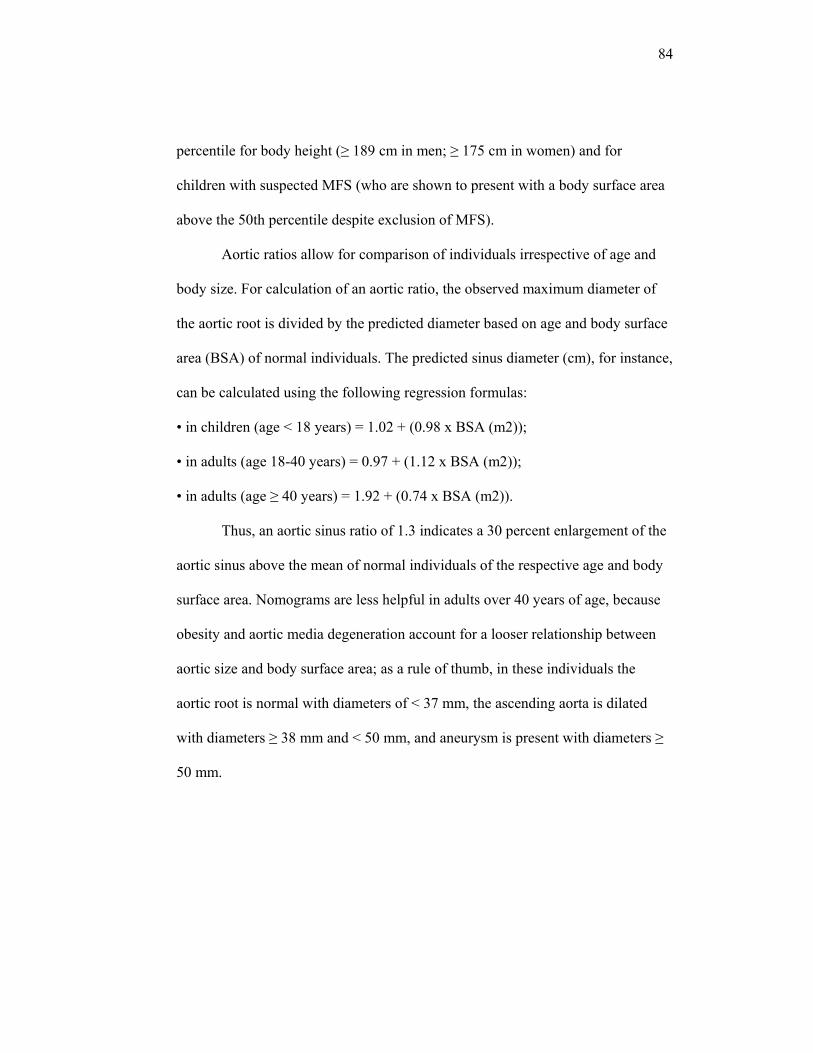
84
percentile for body height (≥ 189 cm in men; ≥ 175 cm in women) and for
children with suspected MFS (who are shown to present with a body surface area
above the 50th percentile despite exclusion of MFS).
Aortic ratios allow for comparison of individuals irrespective of age and
body size. For calculation of an aortic ratio, the observed maximum diameter of
the aortic root is divided by the predicted diameter based on age and body surface
area (BSA) of normal individuals. The predicted sinus diameter (cm), for instance,
can be calculated using the following regression formulas:
• in children (age < 18 years) = 1.02 + (0.98 x BSA (m2));
• in adults (age 18-40 years) = 0.97 + (1.12 x BSA (m2));
• in adults (age ≥ 40 years) = 1.92 + (0.74 x BSA (m2)).
Thus, an aortic sinus ratio of 1.3 indicates a 30 percent enlargement of the
aortic sinus above the mean of normal individuals of the respective age and body
surface area. Nomograms are less helpful in adults over 40 years of age, because
obesity and aortic media degeneration account for a looser relationship between
aortic size and body surface area; as a rule of thumb, in these individuals the
aortic root is normal with diameters of < 37 mm, the ascending aorta is dilated
with diameters ≥ 38 mm and < 50 mm, and aneurysm is present with diameters ≥
50 mm.
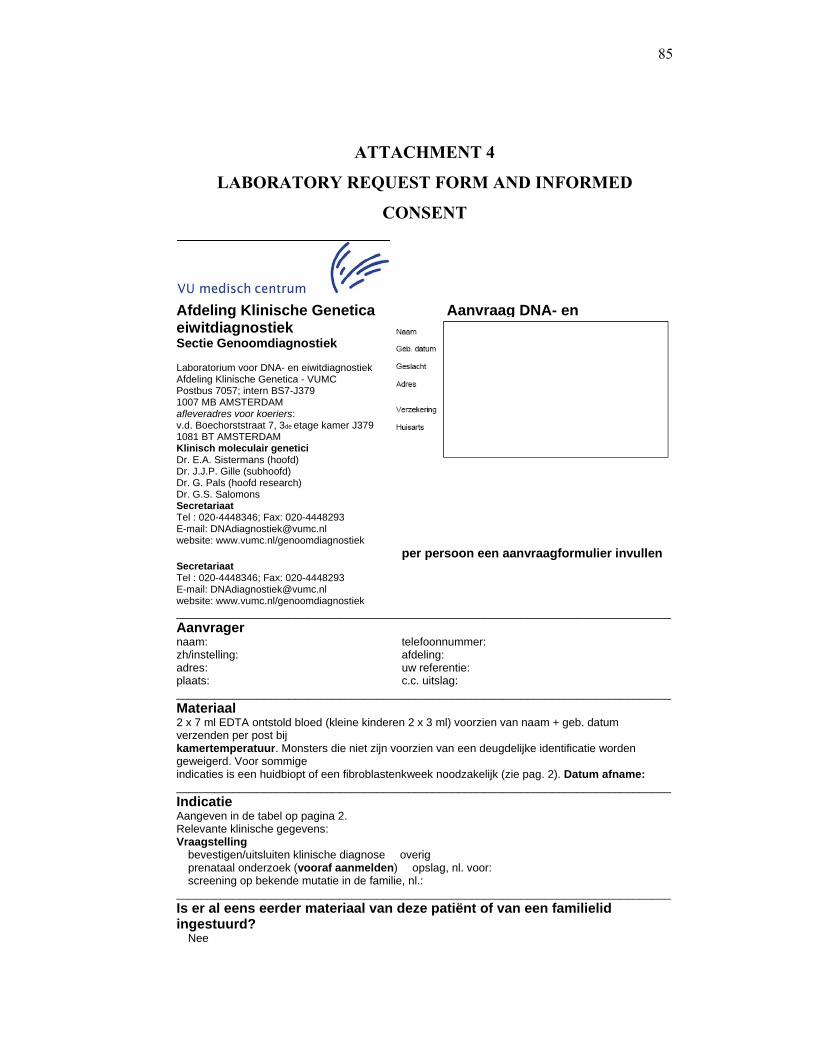
85
ATTACHMENT 4
LABORATORY REQUEST FORM AND INFORMED
CONSENT
Afdeling Klinische Genetica Aanvraag DNA- en eiwitdiagnostiek Sectie Genoomdiagnostiek Laboratorium voor DNA- en eiwitdiagnostiek Afdeling Klinische Genetica - VUMC Postbus 7057; intern BS7-J379 1007 MB AMSTERDAM afleveradres voor koeriers: v.d. Boechorststraat 7, 3de etage kamer J379 1081 BT AMSTERDAM Klinisch moleculair genetici Dr. E.A. Sistermans (hoofd) Dr. J.J.P. Gille (subhoofd) Dr. G. Pals (hoofd research) Dr. G.S. Salomons Secretariaat Tel : 020-4448346; Fax: 020-4448293 E-mail: [email protected] website: www.vumc.nl/genoomdiagnostiek
per persoon een aanvraagformulier invullen Secretariaat Tel : 020-4448346; Fax: 020-4448293 E-mail: [email protected] website: www.vumc.nl/genoomdiagnostiek _______________________________________________________________________________Aanvrager naam: telefoonnummer: zh/instelling: afdeling: adres: uw referentie: plaats: c.c. uitslag: _______________________________________________________________________________Materiaal 2 x 7 ml EDTA ontstold bloed (kleine kinderen 2 x 3 ml) voorzien van naam + geb. datum verzenden per post bij kamertemperatuur. Monsters die niet zijn voorzien van een deugdelijke identificatie worden geweigerd. Voor sommige indicaties is een huidbiopt of een fibroblastenkweek noodzakelijk (zie pag. 2). Datum afname: _______________________________________________________________________________Indicatie Aangeven in de tabel op pagina 2. Relevante klinische gegevens: Vraagstelling � bevestigen/uitsluiten klinische diagnose � overig � prenataal onderzoek (vooraf aanmelden) � opslag, nl. voor: � screening op bekende mutatie in de familie, nl.: _______________________________________________________________________________Is er al eens eerder materiaal van deze patiënt of van een familielid ingestuurd? � Nee
86
� Ja, nl. naam: geb. datum: ref. nr. Stamboom (eventueel aparte stamboom meesturen): � Betrokkene geeft geen toestemming voor anoniem gebruik van lichaamsmateriaal voor research (zie 5.3 op pag. 3). _______________________________________________________________________________In te vullen door het laboratorium ZIS-nr.: familienummer: VD-nummer aanwezig materiaal: ontvangen materiaal: paraaf staflid: Indicaties voor DNA-onderzoek � Achondroplasie (FGFR3) � Alzheimer � PSEN1 � PSEN2 � APP � Apert syndroom � Azoöspermie/oligospermie (CFTR) � Azoöspermie/oligospermie (AZFa/b/c deleties) � Basaal Cel Nevus syndroom (PTCH) � Birt-Hogg-Dubé syndroom (FLCN) � Blackfan-Diamond anemie (RPS19) � Borst- en ovariumkanker � BRCA1 � BRCA2 � BPES (Blepharophimosis, ptosis, en epicanthuis inversus syndroom; FOXL2) � CBAVD (CFTR) � Chorea, erfelijke benigne (TITF1) � Craniosynostose (FGFR2, TWIST) � Crouzon syndroom � Cystic fibrosis (CFTR) � Darmkanker, Lynch syndroom � MLH1 � MSH2 � MSH6 � Darmkanker, MUTYH geassocieerde adenomateuze polyposis � DiGeorge syndroom (22q11-deletie) � Ehlers-Danlos syndroom � COL3A1 (fibroblastenkweek of huidbiopt nodig) � COL5A1 (fibroblastenkweek of huidbiopt nodig) � Elastine (ELN) � Fanconi anemie (alleen na overleg) � Fragiele X syndroom (FRAXA) � Frontotemporale dementie � MAPT � PGRN � CHMP2B � Gorlin syndroom (PTCH) � Hyperferritinemie-cataract syndroom (FTH1) � Hypochondroplasie (FGFR3) � Langer mesomele dysplasie (SHOX) � Loeys-Dietz syndroom � TGFBR1
� TGFBR2 � Marfan syndroom � FBN1 � TGFBR2 � Maternale contaminatie � MLPA microdeletie syndromen (o.a. 22q11 en Williams syndr.) � MLPA subtelomeren � Obesitas (MC4R) � Osteogenesis imperfecta � COL1A1 � COL1A2 � Parkinson, ziekte van � Parkin (Park2) � DJ-1 (Park7) � Pink1 (Park6) � SNCA (Park4) � LRRK2 (Park8) � Pelizaeus-Merzbacher, ziekte van (PLP1) � Pelizaeus-Merzbacher-like disease, autosomaal recessief (GJA12) � Peutz-Jeghers syndroom (STK11) � Pfeiffer syndroom (FGFR2, FGFR3) � Porencephalie (COL4A1) � Prematuur ovarieel falen (FMR1 premutaties) � Pulmonale arteriële hypertensie, idiopatische (BMPR2) � Schmid dysplasie (COL10A1) � Saethre-Chotzen syndroom (FGFR3/TWIST) � Surfactant proteïne B deficiëntie (SFTPB) � Thanatofore dysplasie (FGFR3) � Uniparentale disomie (UPD) � Van de Woude syndroom (IRF6) � Andere indicatie (alleen na telefonisch overleg) Overig DNA-onderzoek Onderzoek dat uitsluitend kan worden aangevraagd na overleg met prof. dr. M.S. van der Knaap, kinderneuroloog ([email protected]) � Megalencephalic leukoencephalopathy with subcortical cysts (MLC1) � Leukoencephalopathie with vanishing white matter (VWM) � Leukoencephalopathy with brain stem and spinal cord involvement and lactate elevation (LBSL)
87
Onderzoek dat wordt verricht binnen het Metabool Laboratorium van het VUmc (Dr. G.S. Salomons) Hiervoor een ander formulier gebruiken dat kan worden aangevraagd bij [email protected] • Alexander, ziekte van (GFAP) • Canavan, ziekte van (ASPA) • Cerebraal creatine deficiëntie syndroom (AGAT, GAMT, SLC6A8) • D-2-hydroxyglutaric dehydrogenase deficiëntie (D2HGDH) • Epilepsie, pyridoxine afhankelijke (ALDH7A1 • GABA metabolisme (ALDH5A1, SSADH, GABA-T) • Glutaryl-CoA dehydrogenase deficiëntie (GCDH) • Homocysteine metabolisme (CBS, MTHFR,MMACHC) • L-2-hydroglutaric dehydrogenase deficiëntie, (L2HGDH) • Malonyl CoA decarboxylase deficiëntie (MLYCD) • Ribose-5-phosphate isomerase deficiëntie (RPI) • Tarui, ziekte van (PFKM) • Transaldolase deficiëntie (TALDO) • X-gebonden creatine transporter defect (SLC6A8) Indicaties voor eiwitonderzoek � Fibroblastenkweek voor enzymonderzoek elders* � Osteogenesis imperfecta, type ___* � Ehlers-Danlos syndroom type ___* � Primaire Ciliaire Dyskinesie/Kartagener syndroom (respiratoir epitheelbiopt nodig) * hiervoor is inzending van een fibroblastenkweek of een huidbiopt noodzakelijk Huidbiopten Afname: • huidbiopten onder steriele condities afnemen, na desinfectie met 70% alcohol (geen jodiumtinctuur) bij voorkeur aan de binnenkant van de onderarm of tijdens een operatie van de randen van de incisieplaats. • Het biopt opvangen in steriel kweekmedium (op verzoek kan dit toegezonden worden). Alleen in noodgevallen een steriele fysiologische zoutoplossing gebruiken. • Indien buiten normale laboratoriumwerktijden een biopt moet worden afgenomen, het materiaal bewaren bij kamertemperatuur (niet op ijs) en de volgende werkdag versturen. Verzending • het materiaal bij voorkeur op maandag, dinsdag of uiterlijk woensdag inzenden per TPG post. Op andere dagen alleen via een koerier. • het materiaal goed inpakken ter bescherming tegen breuk en forse temperatuurdalingen. • op het pakje vermelden “breekbaar” en “bewaren bij kamertemperatuur”. 1. Aanvragen 1.1. Om fouten en vertragingen te vermijden behoren aanvragen op een duidelijke en ondubbelzinnige wijze te worden ingediend. Door gebruik te maken van dit aanvraagformulier komen alle gewenste gegevens aan de orde. 1.2. Met de acceptatie van een aanvraag verplicht de laboratorium zich tot het met zorg en vakmanschap uitvoeren van de gevraagde werkzaamheden volgens de voor de laboratorium geldende kwaliteitscriteria. 1.3. Aanvragen kunnen worden geweigerd indien deze onvoldoende gegevens bevatten om een resultaat te kunnen bereiken dat voldoet aan de geldende kwaliteitscriteria. 1.4. Het laboratorium moet in de gelegenheid gesteld te worden om met de aanvrager/behandelaar te kunnen overleggen over het gevraagde onderzoek. 1.5. De aanvrager wordt verzocht om alvorens patiëntenmateriaal in te sturen, na te gaan of de betreffende patiënt is verzekerd voor klinisch genetische zorg. Indien na uitvoering van een verrichting de patiënt niet verzekerd blijkt, wordt de rekening naar de patiënt gestuurd. 2. Monsters 2.1. De aanvrager levert de te onderzoeken monsters aan bij het laboratorium, voorzien van een deugdelijke identificatie (naam en geboortedatum) en een volledig ingevuld aanvraagformulier. 2.2. Per patiënt 2 x 7 ml EDTA bloed afnemen in onbreekbare buizen (geen glazen buizen), bij kleine kinderen 2 x 3 ml, en per post opsturen bij kamertemperatuur. 2.3. Indien niet wordt voldaan aan het gestelde in 2.1 en 2.2 is het laboratorium niet gehouden het ingestuurde monster in ontvangst te nemen. 2.4. Voor zover bij de indiening van de aanvraag daarover niets is overeengekomen, zal het laboratorium de monsters, c.q. de restanten daarvan na onderzoek, overeenkomstig de eigen voorschriften voor onbepaalde tijd bewaren. 2.5. Alle handelingen en opslag voorafgaand aan de in ontvangstname van een monster vallen buiten de verantwoordelijkheid van het laboratorium.
88
3. Resultaten 3.1. Resultaten in de vorm van onderzoeksuitslagen, adviezen, informatie of welke andere vorm dan ook, worden door het laboratorium in schriftelijke vorm aangeleverd. 3.2. Resultaten komen doorgaans beschikbaar binnen: • Prenataal onderzoek: 2-3 weken • Presymptomatisch / dragerschapbepaling / bevestiging diagnose (bekende mutatie): 6-8 weken • Mutatie scanning (opsporen van nog onbekende mutatie): 3-6 maanden. In geval van spoed kunnen in overleg andere uitslagtermijnen worden afgesproken. 4. Geheimhouding 4.1. Geheimhouding van gegevens is gewaarborgd en vastgelegd in de ziekenhuisvoorschriften van het VU medisch centrum (zwijgplicht over patiëntengegevens). 5. Gebruik patiëntenmateriaal 5.1. Het laboratorium bewaart het verkregen DNA monster van de patiënt voor onbepaalde tijd tenzij een schriftelijk verzoek om het monster te vernietigen is ontvangen van de patiënt of diens wettelijke vertegenwoordigers. 5.2. Het laboratorium gebruikt herleidbaar geanonimiseerd patiënten materiaal voor verder onderzoek (research) in lijn met de oorspronkelijke diagnostische vraagstelling. In geval dit resulteert in voor de patiënt relevante bevindingen zal deze via de oorspronkelijke aanvrager worden geïnformeerd. 5.3. Voor het ontwikkelen van nieuwe en het verbeteren van bestaande technieken gebruikt het laboratorium herleidbaar geanonimiseerd patiëntenmateriaal, o.a. voor controles en validatie. Het laboratorium verzoekt de aanvrager de patiënt hierover te informeren. Mocht deze bezwaar maken tegen het anoniem gebruik van lichaamsmateriaal, dan kan dit op pagina 1 van het aanvraagformulier worden aangegeven.
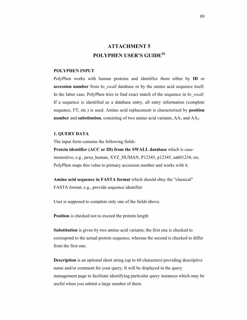
89
ATTACHMENT 5
POLYPHEN USER’S GUIDE26
POLYPHEN INPUT
PolyPhen works with human proteins and identifies them either by ID or
accession number from hs_swall database or by the amino acid sequence itself.
In the latter case, PolyPhen tries to find exact match of the sequence in hs_swall.
If a sequence is identified as a database entry, all entry information (complete
sequence, FT, etc.) is used. Amino acid replacement is characterised by position
number and substitution, consisting of two amino acid variants, AA1 and AA2.
1. QUERY DATA
The input form contains the following fields:
Protein identifier (ACC or ID) from the SWALL database which is case-
insensitive, e.g., pexa_human, XYZ_HUMAN, P12345, p12345, aah01234, etc.
PolyPhen maps this value to primary accession number and works with it.
Amino acid sequence in FASTA format which should obey the "classical"
FASTA format, e.g., provide sequence identifier
User is supposed to complete only one of the fields above.
Position is checked not to exceed the protein length
Substitution is given by two amino acid variants; the first one is checked to
correspond to the actual protein sequence, whereas the second is checked to differ
from the first one.
Description is an optional short string (up to 60 characters) providing descriptive
name and/or comment for your query. It will be displayed in the query
management page to facilitate identifying particular query instances which may be
useful when you submit a large number of them.

90
2. OPTIONS
Structural database (PDB/PQS)
PolyPhen can use two protein structure databases, PDB and PQS. In general,
queries against PDB can be faster than those against PQS. However, use of PQS
(default) is strongly recommended if a user is concerned with residue contacts,
especially inter-subunit.
Sort hits by (Identity/E-value)
Hits are sorted according to the sequence identity or E- value (default) of the
sequence alignment with the input protein.
Map to mismatch (No/Yes)
By default, a hit is rejected if its amino acid at the corresponding position differs
from the amino acid in the input sequence. Mapping to mismatching amino acid
residue should be used with caution only when a protein with known structure and
matching amino acid can not be found.
Calculate structural parameters (For first hit only/For all hits)
In some cases a user may want to check the conservation of structural parameters
of a residue in all hits. By default, parameters are calculated for the first hit only,
since they are expected to be very close in all homologous structures.
Calculate contacts (For first hit only/For all hits)
Contrary to the structural parameters, contacts are by default calculated for all
found hits with known structure. This is essential for the cases when several
PDB(PQS) entries correspond to one protein, but carry different information
about complexes with other macromolecules and ligands (for example, see Fig.2
in [Sunyaev et al 2001])
Minimal alignment length (integer number, default: 100)
PolyPhen will filter out hits with structure whose alignment length with the query
sequence is smaller than the given value.
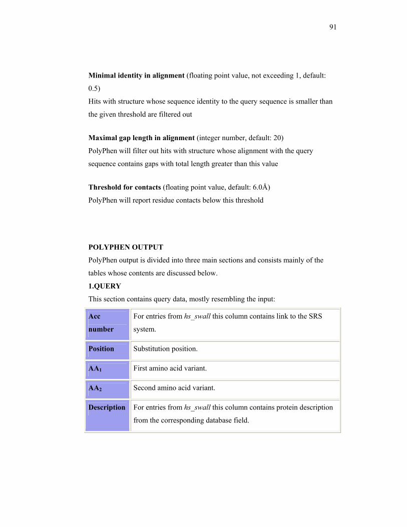
91
Minimal identity in alignment (floating point value, not exceeding 1, default:
0.5)
Hits with structure whose sequence identity to the query sequence is smaller than
the given threshold are filtered out
Maximal gap length in alignment (integer number, default: 20)
PolyPhen will filter out hits with structure whose alignment with the query
sequence contains gaps with total length greater than this value
Threshold for contacts (floating point value, default: 6.0Å)
PolyPhen will report residue contacts below this threshold
POLYPHEN OUTPUT
PolyPhen output is divided into three main sections and consists mainly of the
tables whose contents are discussed below.
1.QUERY
This section contains query data, mostly resembling the input:
Acc
number
For entries from hs_swall this column contains link to the SRS
system.
Position Substitution position.
AA1 First amino acid variant.
AA2 Second amino acid variant.
Description For entries from hs_swall this column contains protein description
from the corresponding database field.
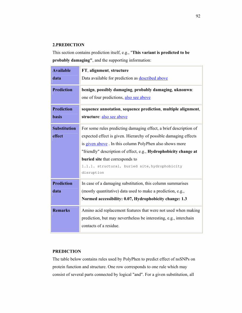
92
2.PREDICTION
This section contains prediction itself, e.g., "This variant is predicted to be
probably damaging", and the supporting information:
Available
data
FT, alignment, structure
Data available for prediction as described above
Prediction benign, possibly damaging, probably damaging, uknonwn:
one of four predictions, also see above
Prediction
basis
sequence annotation, sequence prediction, multiple alignment,
structure: also see above
Substitution
effect
For some rules predicting damaging effect, a brief description of
expected effect is given. Hierarchy of possible damaging effects
is given above . In this column PolyPhen also shows more
"friendly" description of effect, e.g., Hydrophobicity change at
buried site that corresponds to 1.1.1. structural, buried site,hydrophobicity
disruption
Prediction
data
In case of a damaging substitution, this column summarises
(mostly quantitative) data used to make a prediction, e.g.,
Normed accessibility: 0.07, Hydrophobicity change: 1.3
Remarks Amino acid replacement features that were not used when making
prediction, but may nevertheless be interesting, e.g., interchain
contacts of a residue.
PREDICTION
The table below contains rules used by PolyPhen to predict effect of nsSNPs on
protein function and structure. One row corresponds to one rule which may
consist of several parts connected by logical "and". For a given substitution, all
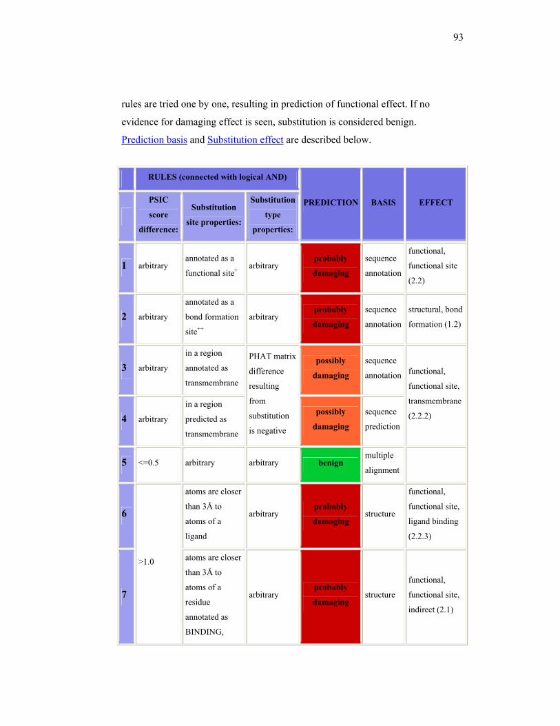
93
rules are tried one by one, resulting in prediction of functional effect. If no
evidence for damaging effect is seen, substitution is considered benign.
Prediction basis and Substitution effect are described below.
RULES (connected with logical AND)
PREDICTION BASIS EFFECT
PSIC
score
difference:
Substitution
site properties:
Substitution
type
properties:
1 arbitrary annotated as a
functional site+ arbitrary
probably
damaging
sequence
annotation
functional,
functional site
(2.2)
2 arbitrary
annotated as a
bond formation
site++
arbitrary probably
damaging
sequence
annotation
structural, bond
formation (1.2)
3 arbitrary
in a region
annotated as
transmembrane
PHAT matrix
difference
resulting
from
substitution
is negative
possibly
damaging
sequence
annotation functional,
functional site,
transmembrane
(2.2.2) 4 arbitrary
in a region
predicted as
transmembrane
possibly
damaging
sequence
prediction
5 <=0.5 arbitrary arbitrary benign multiple
alignment
6
>1.0
atoms are closer
than 3Å to
atoms of a
ligand
arbitrary probably
damaging structure
functional,
functional site,
ligand binding
(2.2.3)
7
atoms are closer
than 3Å to
atoms of a
residue
annotated as
BINDING,
arbitrary probably
damaging structure
functional,
functional site,
indirect (2.1)
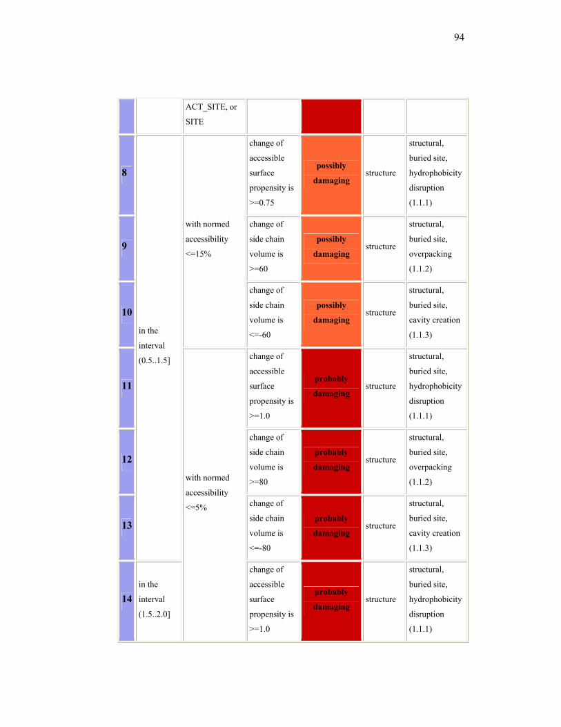
94
ACT_SITE, or
SITE
8
in the
interval
(0.5..1.5]
with normed
accessibility
<=15%
change of
accessible
surface
propensity is
>=0.75
possibly
damaging structure
structural,
buried site,
hydrophobicity
disruption
(1.1.1)
9
change of
side chain
volume is
>=60
possibly
damaging structure
structural,
buried site,
overpacking
(1.1.2)
10
change of
side chain
volume is
<=-60
possibly
damaging structure
structural,
buried site,
cavity creation
(1.1.3)
11
with normed
accessibility
<=5%
change of
accessible
surface
propensity is
>=1.0
probably
damaging structure
structural,
buried site,
hydrophobicity
disruption
(1.1.1)
12
change of
side chain
volume is
>=80
probably
damaging structure
structural,
buried site,
overpacking
(1.1.2)
13
change of
side chain
volume is
<=-80
probably
damaging structure
structural,
buried site,
cavity creation
(1.1.3)
14 in the
interval
(1.5..2.0]
change of
accessible
surface
propensity is
>=1.0
probably
damaging structure
structural,
buried site,
hydrophobicity
disruption
(1.1.1)
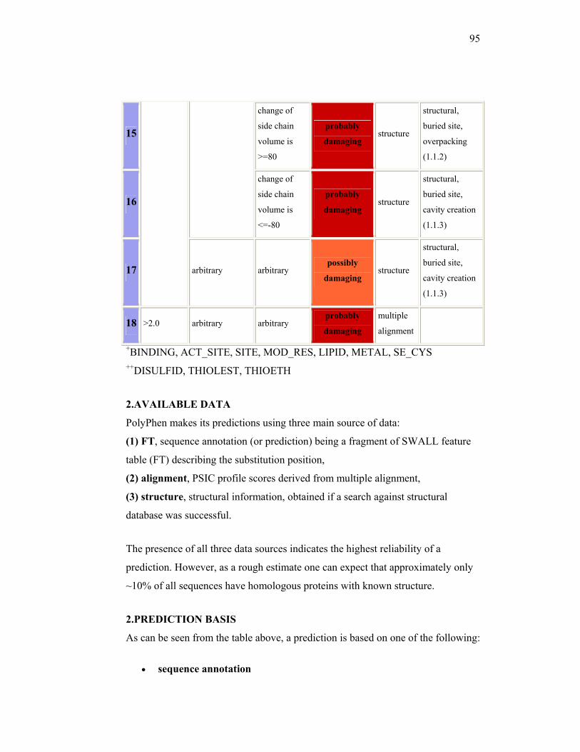
95
15
change of
side chain
volume is
>=80
probably
damaging structure
structural,
buried site,
overpacking
(1.1.2)
16
change of
side chain
volume is
<=-80
probably
damaging structure
structural,
buried site,
cavity creation
(1.1.3)
17 arbitrary arbitrary possibly
damaging structure
structural,
buried site,
cavity creation
(1.1.3)
18 >2.0 arbitrary arbitrary probably
damaging
multiple
alignment
+BINDING, ACT_SITE, SITE, MOD_RES, LIPID, METAL, SE_CYS ++DISULFID, THIOLEST, THIOETH
2.AVAILABLE DATA
PolyPhen makes its predictions using three main source of data:
(1) FT, sequence annotation (or prediction) being a fragment of SWALL feature
table (FT) describing the substitution position,
(2) alignment, PSIC profile scores derived from multiple alignment,
(3) structure, structural information, obtained if a search against structural
database was successful.
The presence of all three data sources indicates the highest reliability of a
prediction. However, as a rough estimate one can expect that approximately only
~10% of all sequences have homologous proteins with known structure.
2.PREDICTION BASIS
As can be seen from the table above, a prediction is based on one of the following:
• sequence annotation

96
• sequence prediction
• multiple alignment
• structure
depending on the rule used to make it.
97
ATTACHMENT 6
SIFT USER GUIDE27
SIFT takes a query sequence and uses multiple alignment information to predict tolerated and deleterious substitutions for every position of the query sequence. SIFT is a multistep procedure that (1) searches for similar sequences, (2) chooses closely related sequences that may share similar function to the query sequence , (3) obtains the alignment of these chosen sequences, and (4) calculates normalized probabilities for all possible substitutions from the alignment. Positions with normalized probabilities less than 0.05 are predicted to be deleterious, those greater than or equal to 0.05 are predicted to be tolerated. Procedure (the details):
1. Get related sequences. A PSI-BLAST search against a database is executed on the query sequence. Parameters: 4 iterations, expectation value .0001, e-value threshold for inclusion in multipass model 0.002 Update 05/15/01 Number of PSI-BLAST iterations reduced to 2 to save time and prevent the search from diverging.
2. Choose closely related sequences. As described in Genome Research 11:963-87: We desire to have sequences that are similar in function as well as structure to the query sequence. To do so, we select only a subset of sequences from the PSI-BLAST results.
a. Group sequences found from the PSI-BLAST search that are more than 90% identical together and make a consensus sequence for each group by choosing the amino acid that occurs most frequently at each position.
b. MOTIF finds conserved regions among the query sequence and the consensus sequences from (a) that were derived from at least two sequences.
c. After the conserved regions in the query sequence have been identified by MOTIF, these regions are extracted from the sequences aligned by PSI-BLAST.
d. The conserved regions of the query sequence and those consensus sequences more than 90% identical are converted to a PSI-BLAST checkpoint file.
e. The checkpoint file is given to PSI-BLAST to search among the remaining conserved regions of the consensus sequences not included in the seed checkpoint file. The top hit is added to the alignment corresponding tothe seed checkpoint file and the conservation over the entire alignment of conserved regions is calculated. If conservation does not decrease, the
98
consensus sequence is added to the alignment and the checkpoint file rebuilt. (e) iterates until conservation decreases.
OR
SIFT by conservation: In the original version of SIFT, an arbitrary number of sequences is added. In this version, sequences are continually added until they reach a sequence conservation cutoff, set by the user. If the sequences for which prediction is based on are very diverse (low conservation cutoff), only substitutions at the strongly conserved positions will be predicted as deleterious. If the sequences chosen for prediction are very similar to each other (high conservation cutoff), then most substitutions will be predicted as deleterious. Users can choose the degree of sequence conservation: they can opt for detecting most of the deleterious substitutions (use a high sequence conservation) , or predict fewer deleterious substitutions but with a high level of certainty (use a low sequence conservation).
f. Group sequences found from the PSI-BLAST search (step 1) that are more than 90% identical together and make a consensus sequence for each group by choosing the amino acid that occurs most frequently at each position.
g. The query sequence and its checkpoint file is given to PSI-BLAST to search among the consensus sequences. The top hit is added and aligned to the query sequence. Information is calculated for each position in the alignment, and the median of these values is obtained. If the median conservation over all positions does not fall below a given cuttoff, the hit is retained in the alignment and the checkpoint file rebuilt. The process repeats until the median conservations as long as the median information does not fall below the cutoff.
The sequences picked from this iterative procedure are chosen as closely related sequences. You can also submit your own sequences.
3. Obtain alignment. Since PSI-BLAST alignments are fairly accurate and long (Sauder & Dunbrack, 2000), we obtain the alignment of the sequences chosen in (2) from the initial PSI-BLAST search results (1). You can also submit your own alignment of your query sequence with other sequences.
4. Calculate probabilities. At each position of the alignment, each amino acid i appears at a frequency ni. Using the ni's, the probabilities of amino acids are estimated according to Dirichlet mixtures (di's. The final probability of an amino acid appearing at a position, pi, is a weighted average of the observed
99
frequencies and the Dirichlet estimation. The weight of the observed frequencies is the number of sequences used to construct the alignment. The weight of the Dirichlet estimated probabilities is an exponential function of a diversity measure (Div) calculated by
Div = SUM ( ranki * ni)
where ranki is the rank amino acid i has in reference to the original amino acid when BLOSUM62 substitution scores for the original amino acid are ranked from highest to lowest. Probabilities are normalized by dividing by max{Pr(amino acid)}. Update: 08/08/01: Prior to calculating the probabilities, sequences > 90% identical to the query sequence are removed. This eliminates the possibility that the sequence containing your substitution of interest is already represented in the database therefore and will trivially be predicted as tolerated.
• We have found by comparison to experimental data that substitutions with less than 0.05 are deleterious. We use this as a cutoff for prediction. We strongly suggest users examine the normalized probabilities manually. If your substitutions are slightly above the 0.05 cutoff, you might want to consider this as a deleterious substitution.