klarifikasi: (lampiran bukti kehadiran)
TRANSCRIPT
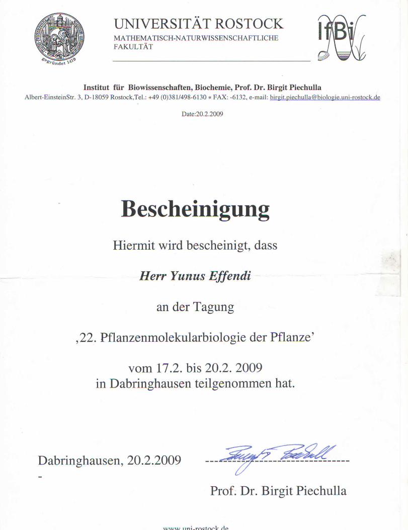
Klarifikasi: (lampiran bukti kehadiran)
Poster yang berjudul: “Lebensfahige Mutante des Auxin-Bildungs-Proteins 1 (ABP1) zeigen
morphologische merkmale ung langsamere regulation von IAA-genen“ dipresentasikan dalam
bentuk poster presentation pada konferensi tahunan tentang 22nd Molecular Biology of The Plant
(22. Tagung Molekularebiologie der Planzen), 17-20 Februari 2009. Penulis sebagai first author
pada presentasi poster ini. Konferensi tersebut diselenggarakan secara rutin di kota Dabringhausen
Jerman dengan sponsor utama Deutsche Botanische Gesellschaft (DBG). Pada 22 Tagung
(Conference 22nd) koordinator untuk kegiatan saat itu adalah Universitaet Rostock – Jerman (Prof.
Dr. Birgit Piechulla sebagai Koordinator konferensi). Konferensi ini tidak menyediakan adanya
proseding sehingga publikasi ini tergolong tidak dimuat dalam prosiding. Informasi tentang
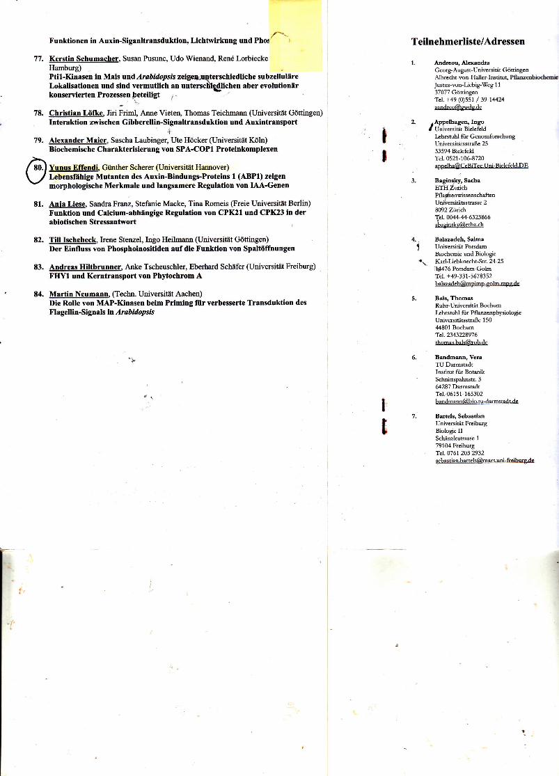
konferensi (penyelenggara, peserta dan poster/makalah) terinfokan pada abstract book
(dilampirkan). Sebagai bukti keikutsertaan kami dilampirkan
Sertifikat kehadiran
Artikel terkait poster yang dipresentasikan
scan Abstract book (info Organizing Committee)
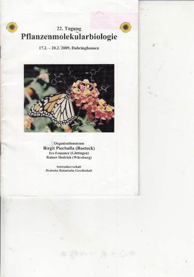
22. Tagung
P fl a n zenmo le kul a rb i olo gi e
17.2. - 20.2.2009, Dabringhausen
e.:f'.f ry'
f" t:.:5r:
Organisationsteam
Birgit Piechulla (Rostock)Ivo Feussner (Giittingen)
Rainer Hedrich (Wiirzburg)
SchirmherrschaftDeutsche Botanische Gesellschaft
77.
78.
Funktionen in Auxin-Siganltransduktion, Licbtwirkung und Phos ft',
Kerstin Schumacher, Susan Pusunc, Udo Wienand, Rend LorbieckeHamburg)Ptil-Kinasen in Mais rnd Arabidopsis zeige*pqterschiedliche subzellulireLokrlisationen und sind vermutlich an untersch\lichen aber evolutionirkonserviertenProzessenleteiligt i-
Chrlstian LOIke, firi irimt, Anne Vieten, Thomas Teichmann (Universitat Gdttingen)Interaktion zwischen Gibberellin-Signaltransduktion und Auxintransport
IAlexander Maier, Sascha Laubinger, Ute Hticker (Universitiit Kdln)Biochemische Charakterisierung von SPA-COPI Proteinkomplexen
Yunus Effendl Gibrther Scherer (Universitit Hannover)Lebensflhige Mutanten des Auxin-Bindungs-Proteins I (ABPI) zeigenmorphologische Merkmale und langsamere Regulation von IAA-Genen
Ania Liese, Sandra Franz, Stefanie Macke, Tina Romeis (Freie Universitiit Berlin)Funktion und Calcium-abhlngige Regulation von CPK2I und CPK23 in derabiotischen Stressantwort
TiIl Ischebeck, Irene Stenzel, Ingo Heihnaon (Universiuit Gdttingen)Der Einfluss von Phosphoinositiden aufdie Funktion von Spaltiiffnungen
Andreas Hiltbrunner, Anke Tscheuschler, Eberhard Schiifer (Universitat Freiburg)FIIYI und Kerntransport von Phytochroim A
Martin Neumann, (Techn. Universit6t Aachen)Die Rolle von MAP-Kinasen beim Priming fiir verbesserte Transduktion des
Flagellin-Signals h Arabidopsis
79.
o81.
82.
E3.
E4.
II
Teilnehmerliste/Adressen
1. Andreou, AlxandraGeorg-August-Universitiit G6niogmAlbrecht-von-Haller-Instimt, Pfl ambioctoiJustus-Yon-Liebig-Iweg 1 1
37077 GcittingmTel. +49 (0)551 / 39-14424amdreo@,gwdg.de
2. .Appelbagen, IngoI Univemitiit Bielefeld
I:hrstuhl fiir GenomfonchungUniversititsstraBe 25
33594 BielefeldTel. O52l-106-8720
3. Bagins-ky, Sachap.Ttt Z*i"hPfla{mwissenschaftenUo&ersit4tsstasse 2B(h2zftic}-\e1.004+4{[email protected]
4. Balazadch, Salma
! UniversititPotsdmBiochemie md Biologie
"\ Kad-Uebknecht-Str.2+25. i!4476 Potsdm-Golrn
aal. +49-331-5678152
Bals, ThomasRuhr-Universitdt Bochmkhrstuhl fiir PflanzenphysiologieUniversitiissttaBe 1 50,{4801 [email protected]
Bandmmn,VemTU DumstzdtInstitut fiir BotmikSchnittspahnstr. 364287 DtrmstadtTel. 06151-165302
Bartels, SebastimUniversitit FreibugBiologie IISchiinzlesmsse 1
79104 FreibwgaeL 0761 2O3 2932
t,
t
,{'t

Lebensfahige Mutante des Auxin-Bildungs-Proteins 1 (ABP1) zeigen
morphologische merkmale ung langsamere regulation von IAA-genen
Yunus Effendi, Günther F. Scherer
1Leibniz Universität Hannover, Institut für Gartenbauliche Produktionssysteme, Abt. Molekulare
Ertragsphysiologie, Herrenhäuser Str. 2, D-30419 Hannover, Germany
E-Mail: Corresponding: [email protected]
Abstract
Auxin signaling in plant is regulated by TIR1 as ist major receptor through ubiquitination and gene
transcription activities. However, there is remain question about a number of auxin-regulated
enzymatic activities or processes within short time after auxin application, such as activation of
phospholipase A activity (Scherer and Andre, 1989; Paul et al, 1998) after 2 min, regulation of
channel activity within 3 min (Martin et al, 1991; Rueck et al, 1993), and activity of MAP kinase
within 5 min (Mockaitis and Howell, 2000) after auxin application. Those processes can not be
explained via gene transcription activities of TIR1 complex. Auxin Binding Protein 1 (ABP1) is a
candidate for a second auxin receptor, even though ABP1 does not show any similarity with
common receptor and it is located dominantly in the lumen of ER where the pH is to high for auxin
binding (Tien et al, 1995). No pre-report described auxin stimulated gene regulation through ABP1
action. Moreover, since loss-of-function in ABP1 leads to lethality in an early stage of embryo
development (Chen et al, 2002), it is difficult to obtain evidence the involvement of ABP1 in
auxin-dependent development. Because of the embryo lethality from abp1/ABP1 plants only 2:1
mixtures of abp1/ABP1 and wt plants can be obtained.
We analyzed first kanamycin resistant viable heterozygous abp1/ABP1 plants which produce a
progeny mixture 2:1 abp1/ABP1 plants. For investigation on gravitropism, phototropism and
flowering in SD, we used such mixed populations. Despite this drawback, it is obvious that
gravitropism of root and shoot and phototropism of the shoot is defect and early flowering in SD
is clearly indicated. Short day plants also do not show obvious morphological differences to wt
plants. We show differences in expression level of IAA gene family (IAA2, IAA11, IAA13, IAA14,
IAA19 and IAA20) in heterozygous abp1/ABP1 2:1 (selected as kanamycin resistant) and wt
Arabidopsis thaliana (ecotype Wassilewskija). Heterozygous abp1/ABP1 show less fold
expression of those IAA genes in comparison to wt after 0.1 µM auxin treatment (30 min and 60
min). After 30 min auxin application, IAA2, IAA11, IAA13, IAA14, IAA19 and IAA20 genes are
expressed 2,3,2.4, 12, 30, and 5 times more in wt than in heterozygous abp1/ABP1 plants,
respectively (P<0.05). Phenotypic data show that heterozygous abp1/ABP1 plants produce
inflorescence earlier (70 days, SE= 0.46) in comparison to wt (75 days, SE=0.63), fewer rosette
leaves and fewer cauline leaves. Moreover, in comparison to wt, the roots of heterozygous
abp1/ABP1 plants show less responsivity to gravity and the hypocotyls are less responsivity to
gravity and lateral light.
We also are in the process of making plants were transformed with different vector constructs
containing mutated amino acids. By selection and segregation, eventually stable double
homozygous plants can be obtained. Currently, plants still may contain wild type protein and over
express the mutated gene. These plants seem to flower early also in LD as indicated by the number
of rosette and cauline leaves. Early flowering may be the cause of the dwarfed appearance of
mutant 6. A low number of stems may indicate a change in apical dominance. Curly leaves on
almost all mutants are reminiscent of the phenotype of phot1 phot2 double mutant.
Keyword: heterozygous abp1/ABP1, auxin receptor, mutants
INTRODUCTION
Our knowledge of auxin signaling made considerable progress in the last few years. The
identification of the receptor TIR1 and its mechanism of action have provided the crucial missing
component in a pathway that can now be seen to be sufficient to account for how auxin can turn a
gene on. Following decades of research on auxin molecular biology, several elements of the auxin
signaling pathway have been elucidated. These include two or even three types of apparent
receptors that perceive the auxin signal. Functions influenced by these receptors are gene
expression and also possibly cell wall modifications important for cell expansion (Napier et al.,
2001). The apparently two major types of receptors are Auxin Binding Protein 1 (ABP1), a
membrane-bound protein thought to initiate cell wall expansion, and more recently Transport
Inhibitor Response 1 (TIR1) and related auxin signaling F-box (AFB) proteins, soluble proteins
that initiate the regulation of gene expression (Dharmasiri et al., 2005). Recently, intensive
investigation about auxin receptor and its function on mediating the expression of auxin induced
genes is focused on TIR1 and the related auxin signaling F-Box proteins. Therefore, it is more
accepted and explainable that TIR1 is a major auxin receptor. However, some early and rapid
physiological processes which appear after auxin application within less than 30 minutes remain
unexplained by function TIR1 to initiate the regulation via proteolysis of IAA transcription factors
and transcriptional activation of early auxin-induced genes. Because it takes time to generate new
proteins by gene regulation which is inevitably slower than signaling, for example, by membrane
depolarization or ion fluxes, rapid responses to auxin might not be TIR1-mediated. Some are
linked to ABP1 (Badescu and Napier, 2006).
Although currently ABP1 as an auxin receptor is still subject to debate, it has been well known
that some physiological processes in plant are mediated by ABP1. Predominantly, ABP1 is found
in lumen of endoplasmic reticulum where it is retained by a KDEL sequence (Jones and Herman,
1993). The possible functional role of the protein inside the ER is nothing known but there is an
experiment evidence for a function of ABP1 at the outer surface of the plasma membrane (LeBlanc
et al., 1999). The pH estimate of the ER lumen is based on an indirect assay, which indicates that
the pH is closer to pH 7 than to the binding optimum of pH 5.5. These results indicate that ABP1
does not bind auxin within the ER and point to a site of action that is post-ER (Tian et al., 1995).
Klämbt (1990) proposed a plasma membrane docking protein as a model for ABP1 action. He
postulated that a small amount of ABP1 manages to escape KDEL retention and is excreted to the
apoplasmic space. In fact, there is some experimental evidence supporting this idea (Jones and
Herman, 1993; Diekmann et al., 1995). After binding extracellular auxin, ABP1 was postulated to
interact with a hypothetical trans-membrane docking protein which transduces the signal into the
cell. ABP1 has been shown to be located at the plasma membrane using immunocytochemisty in
conjunction with electron microscopy (Jones and Herman, 1993) and silver-enhanced fluorescence
microscopy (Diekmann et al., 1995).

Another type of auxin binding protein, ABP57 was found in rice (Kim et al., 2000). It was has
shown that after IAA application ABP57 also binds directly to plasma membrane H+-ATPases and
stimulates proton extrusion in rice. However, auxin binding proteins (ABPs) appears to be able to
activate the plasma membrane H+-ATPases in the presence of auxin (Steffens et al., 2001). Taken
together, these data indicate that ABP1 binds auxins in a specific and physiological meaningful
manner at the plasma membrane to bring about rapid hormone responses. However, no data exist
to show that ABP1 may trigger gene regulation.
The slower but important receptor-triggered functions in auxin physiology required gene
regulation. Such functions are: adventitious and lateral root development and differentiation,
sustained elongation, frit development, ethylene induction, xylem and phloem differentiation.
Only cell division and, perhaps in part, all elongation have been shown require ABP1 but not these
other functions (Campanoni and Nick, 2005). Gene regulation by an auxin receptor is well
explained by TIR1 and homologous AFB genes and regulation of IAA genes and the heterodimeric
IAA/ARF complexes acting as transcription factors (Parry and Estelle, 2006). Therefore, TIR1 and
the homologous AFB’s are regarded as the functionally major receptors. Especially, because the
influence of ABP1 on gene regulation is unknown it is still an open question how real functional
importance of ABP1 is to be evaluated. Therefore, approaches using mutants of the ABP1 gene
and protein are needed
To elucidate this subject, some efforts have been done by many groups. ABP1 as an auxin receptor
remains mysterious. Nevertheless, recently two different workgroups, who work with ABP1 gene,
described a knockout abp1 mutant (Chen et al., 2001) and the crystallographic structure of ABP1
(Woo et al., 2002), which opened a new perspective for investigating ABP1 as a receptor. Chen et
al. (2001) found that a homozygous abp1 T-DNA-mutated Arabidopsis thaliana is lethal at
embryonic stage. The embryos are arrested at globular stage and form a clump of cells but no plant.
This loss of function which leads to a lethal condition in the plant indicates that ABP1 must be an
essential gene in the plant. Moreover, according Chen et al. (2001) plants with one copy
(heterozygous abp1) can grow as a normal plant. This information shows another difficult thing to
deal with ABP1 gene. Generally, understanding of gene function can be performed by altering or
abolishing the expression of the gene. It can be done by mutating the essential part of the gene by
T-DNA insertion which leads to lost of function of the gene, so then the downstream processes
which relate with this gene can be observed. Unfortunately, in case of ABP1 gene, this technique
cannot be easily performed, because null mutation of ABP1 gene will confer to lethality to the
plant. Making a ‘heterozygous’ T-DNA insertional mutant does not confer lethality on plant. A
solution to the dilemma for getting mutants of the ABP1 gene is to use the viable heterozygous
insertional abp1 mutants to transform them with a mutated cDNA ABP1. Transformed plants then
can be selected which are homozygous for the insertion so that no wild type ABP1 protein is
expressed but only the mutated ABP1 protein. During the course of the work it also became
apparent that plants having an insertion in the ABP1 gene in heterozygous constellation already
have a clear mutant phenotype, so that this was also investigated.
MATERIAL AND METHODS
Plant material
Arabidopsis thaliana wild type and heterozygous abp1 mutant ecotype Wassilewskija (Ws-2) were
used as object plant. Heterozygous abp1 mutant seeds were obtained from Nottingham
Arabidopsis Stock Center (NASC) England. Arabidopsis seeds were grown on compost soil
containing silica sand. Aplants were grown in climate chamber or in greenhouse under long day
condition (16 h light/8 h dark). Pre-sterilized seeds were sowed on 0.5X Murashige and Skoog
medium (Murashige and Skoog, 1962) supplemented with 1% (w/v) sucrose and containing
100µg/ml Kanamycin antibiotic. Plants were grown under 18h white light 22oC condition and
transferred to soil after four primary leaf appear then they were grown in green house under long
day condition (16 h day light/8 h dark cycle).
Phenotypic characterization of the heterozygous abp1 T-DNA mutant
Phenotypical characters were measured from 11 days old plants. The seedlings were grown as
described above. Scanned of the seedlings were then the roots length and waving pattern were
scanned with CanonScan 8800F (resolution 600 dot per inchi). Root length and waving pattern
were measured from ten of both wild type and heterozygous abp1 plants by using AxioVisio LE
Ver.4.6 software (Zeiss-Germany).
Auxin treatment
Three different concentrations of Indole-3-acetic acid (0µM, 0.03µM and 0.1µM) were used and
applied for 60 min, 30 min and 0 min to the plants. Due to the handling of the samples, t = 0 min
in this experiment referred to about 10 second. Dimethylsulfoxid (DMSO) treatments with equal
concentration as much as auxin treatments were used as control. Both of these treatments were
applied to heterozygous abp1 and wild type plants. Two auxin concentrations (0µM and 0.1µM)
were used for quantitative real time PCR experiment with the similar time course as semi-
quantitative experiment. After the seeds were surface sterilized, seeds were grown on LB agar
medium containing 100µg/ml kanamycin. Two days after stratification at 4oC in dark condition,
plates were placed under 18h white light 22oC condition until seedling has 4-6 primary leaf. Then
growth further on 1 ml of 0.5X MS medium containing appropriate IAA concentration was added
to the plates. Plates were incubated at room temperature at given time condition (short time, 30
min and 60 min) with gently shaking. Rapidly plants were dried by placing on clean tissue papers,
stored in 2 ml sterile eppendorf tubes and frozen as soon as possible by dipping on liquid nitrogen.
Samples were placed on -80oC until RNA was extracted.
RNA Isolation and cDNA synthesis
Total RNA was isolated from fresh samples using NucleoSpin RNA Plant kit (Macherey-Nagel).
Isolation of RNA was performed according the manufacture's protocol. A 95l DNase was applied
to each samples reaction mix which was made by mixing 90l reaction buffer for rDNase and 10l
rDNase. The RNA were eluted by adding 60l RNase-free water to the center of the membranes
and then centrifuged 1 min at 11.000x g. Purity and concentration of RNAs were measured using
spectophotometer U 3500 (Hitachi-Japan). RNAs then were stored on -20oC. Complementary
DNA (cDNA) were synthesized by using RevertAidtm H Minus First Strand cDNA Synthesis Kit
(Fermentas). For each samples, a 4-5 g of total RNA was mixed with 1l random hexamer primer
(0.5g/l) in a 200l sterilized PCR tube and added deionized-nuclease free water to final volume
12l (4l 5X reaction buffer, 1l Ribonuclease inhibitor (20U/l) and 2l 10mM dNTP mix, 1l
RevertAidTM H Minus M-MuLV RT (200U/l)). The mixtures then were incubated at 42oC for 60
min and the reaction was stopped by heating at 70oC for 10 min.
Quantitative Real Time PCR and data analysis
The relative amount of gene expression of auxin-induced early genes in wild type and
heterozygous abp1 mutant plants were measured by performing quantitative real time PCR. Four
IAA family genes (IAA2, IAA11, IAA13 and IAA20) were analyzed their expression after auxin
treatment on different time courses application. Quantitative PCR reactions were performed using
1 ll of sixfold diluted cDNA, 200 nM primers and 0.2· Power SYBR Green PCR master mix
(Invitrogen, http://www.invitrogen.com/) in a StepOnePlusTM system (Applied Biosystems,
http://www.appliedbiosystems.com/). For each pair of primers, the threshold value and PCR
efficiency value were determined using cDNA diluted tenfold each time in five dilution steps. For
all primer pairs, including the internal standard gene, 18S rRNA, the PCR efficiency was >99%.
The specificity of PCR amplification was examined by monitoring the presence of a single peak
in the melting curves for quantitative PCR. Amplicons were checked for fragment length on 4%
agarose gels. For each determination, two to three biological repeats and three technical replicates
for each determination were performed for the subsequent PCR reaction. Relative expression was
calculated according to the DDCt method using the equation: relative expression = 2)[DC tsample
) DC tcontrol] , where DCt = Ct(sample gene) ) Ct(reference gene) and Ct refers to the threshold
cycle determined for each gene in the early exponential amplification phase (Livak and
Schmittgen, 2001). The expression level for the control treatment was set as 1-fold. For statistical
analysis, REST 2008 software (Pfaffl et al., 2002) was used.
Gene Construction for mutagenesis
Preparation of ABP1 cDNA
The cDNA of Auxin Binding Protein 1 for this experiment was obtained from Dr. Thomas Reinard
(Institute Plant genetic – Leibniz Universität Hannover). The ABP1 cDNA was consisted of 168
amino acids with Strep - Flag tags on its C terminus and KDEL motif in the end of the cDNA
sequence (figure 6). Before the cDNA was cloned into Entry vector pENTR-D TOPO (Invitrogen),
minor modification was performed by adding stop codon AUG in the end of cDNA sequence.
Amplification of cDNA was carried out with ABP1 specific forward primer (5'-
CACCATGGATGATCGTACTTTC-3') and reverse primer (5'-CCTGAGATC
TCAAGTAGGAAGCGTC-3') with PCR conditions 94oC 4 min, 34 cycles (94oC 30 sec, 54oC 30
sec, 72oC 40sec). The vector was transformed into competent cell Escherichia coli strain TOP 10
(Invitrogen) and cultured in Luria Bertani (LB) medium (see appendices Table 5) supplemented
with 100µg/ml kanamycin under 37oC for 16 hour. Ten colonies were picked up and cultured
further on LB medium containing 100µg/ml kanamycin. Plasmid was isolated after cell cultures
were incubated 37oC with rigorous shaking for 16 hour using SV Miniprep kit (Promega)
according the manufacture's protocol and was used as template for mutagenesis step.
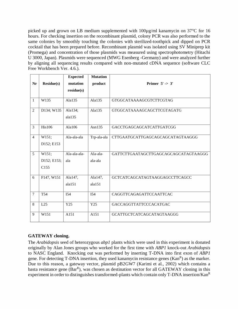
Site-directed mutagenesis
Mutagenesis in this experiment was carried out by using site-directed mutagenesis kits (Invitrogen)
according to the manufacture's protocol. Only one primer for each mutation was applied for each
mutagenesis. Primers for mutagenesis were shown in tabel 1. Mutagenesis was also carried out
according to Zheng et al. (2004) with minor modification. The mutated-vectors were then
transformed into competent E. coli strain TOP 10 or BMH 71S (Promega) using heat shock
method. From each transformation plates (from different mutation constructs), ten colonies were
picked up and grown on LB medium supplemented with 100µg/ml kanamycin on 37oC for 16
hours. For checking insertion on the recombinant plasmid, colony PCR was also performed to the
same colonies by smoothly touching the colonies with sterilized-toothpick and dipped on PCR
cocktail that has been prepared before. Recombinant plasmid was isolated using SV Miniprep kit
(Promega) and concentration of those plasmids was measured using spectrophotometry (Hitachi
U 3000, Japan). Plasmids were sequenced (MWG Esenberg -Germany) and were analyzed further
by aligning all sequencing results compared with non-mutated cDNA sequence (software CLC
Free Workbench Ver. 4.6.).
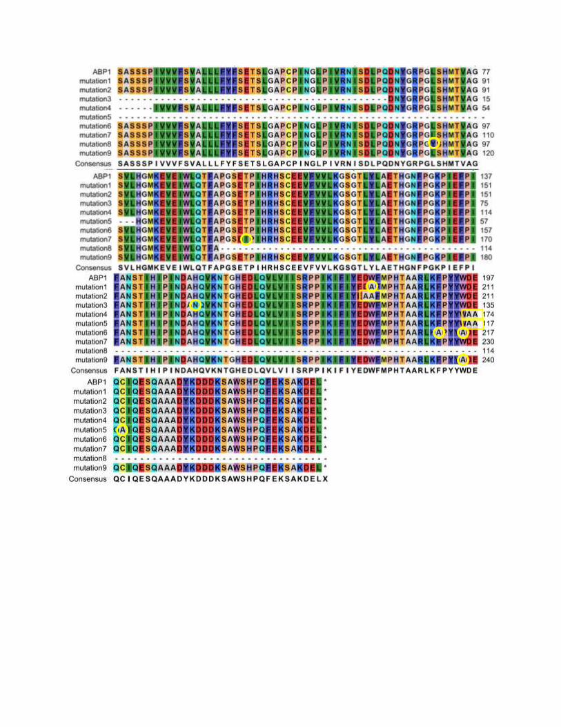
Nr Residue(s)
Expected
mutation
residue(s)
Mutation
product Primer 5' -> 3'
1 W135 Ala135 Ala135 GTGGCATAAAAGCGTCTTCGTAG
2 D134; W135 Ala134;
ala135
Ala135 GTGGCATAAAAGCAGCTTCGTAGATG
3 His106 Ala106 Asn135 GACCTGAGCAGCATCATTGATCGG
4 W151;
D152; E153
Ala-ala-ala Trp-ala-ala CTTGAATGCATTGAGCAGCAGCATAGTAAGGG
5 W151;
D152; E153;
C155
Ala-ala-ala-
ala
Ala-ala-
ala-ala
GATTCTTGAATAGCTTGAGCAGCAGCATAGTAAGGG
6 F147, W151 Ala147,
ala151
Ala147,
ala151
GCTCATCAGCATAGTAAGGAGCCTTCAGCC
7 T54 I54 I54 CAGGTTCAGAGATTCCAATTCAC
8 L25 Y25 Y25 GACCAGGTTATTCCCACATGAC
9 W151 A151 A151 GCATTGCTCATCAGCATAGTAAGGG
GATEWAY cloning.
The Arabidopsis seed of heterozygous abp1 plants which were used in this experiment is donated
originally by Alan Jones groups who worked for the first time with ABP1 knock-out Arabidopsis
to NASC England. Knocking out was performed by inserting T-DNA into first exon of ABP1
gene. For detecting T-DNA insertion, they used kanamycin resistance genes (KanR) as the marker.
Due to this reason, a gateway vector, plasmid pB2GW7 (Karimi et al., 2002) which contains a
basta resistance gene (BarR), was chosen as destination vector for all GATEWAY cloning in this
experiment in order to distinguishes transformed-plants which contain only T-DNA insertion/KanR
(heterozygous abp1 plant) and heterozygous abp1 plants with mutated-ABP1 gene insertion (KanR,
BarR). Recombinant entry vector pENT-D TOPO was ligated with pBGW7 by using Gateway LR
clonase II enzyme mix (Invitrogen). LR reactions were performed by mixing 1-7µl recombinant
entry vector with 150 ng destination vectors and added 2 µl LR clonase II enzyme mixes. Reactions
were incubated overnight at 25oC. One microliter of LR reaction products were transformed into
TOP 10 competent cells and cultured on LB medium containing 100µg/ml spectinomycin and
incubated for 16 hour at 37oC. Ten positive colonies of each constructs were cultured on LB liquid
medium supplemented with 100µg/ml spectinomycin and recombinant plasmids (expression
vector) were isolated for further transformation into Agrobacterium.
RESULTS
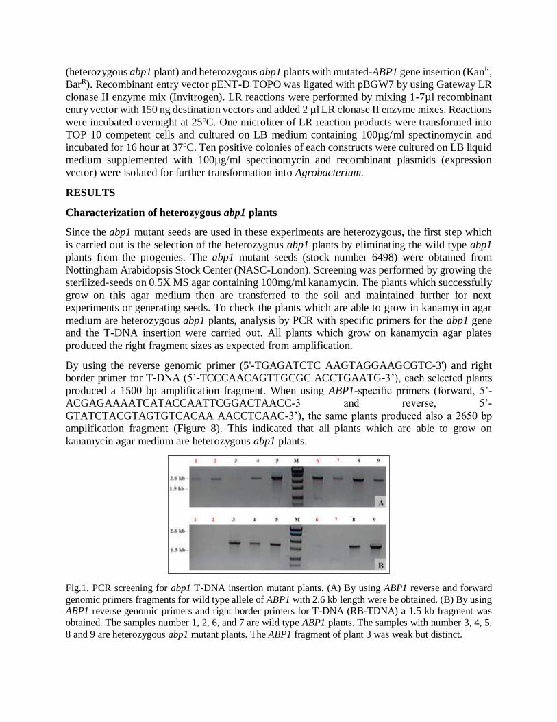
Characterization of heterozygous abp1 plants
Since the abp1 mutant seeds are used in these experiments are heterozygous, the first step which
is carried out is the selection of the heterozygous abp1 plants by eliminating the wild type abp1
plants from the progenies. The abp1 mutant seeds (stock number 6498) were obtained from
Nottingham Arabidopsis Stock Center (NASC-London). Screening was performed by growing the
sterilized-seeds on 0.5X MS agar containing 100mg/ml kanamycin. The plants which successfully
grow on this agar medium then are transferred to the soil and maintained further for next
experiments or generating seeds. To check the plants which are able to grow in kanamycin agar
medium are heterozygous abp1 plants, analysis by PCR with specific primers for the abp1 gene
and the T-DNA insertion were carried out. All plants which grow on kanamycin agar plates
produced the right fragment sizes as expected from amplification.
By using the reverse genomic primer (5'-TGAGATCTC AAGTAGGAAGCGTC-3') and right
border primer for T-DNA (5’-TCCCAACAGTTGCGC ACCTGAATG-3’), each selected plants
produced a 1500 bp amplification fragment. When using ABP1-specific primers (forward, 5’-
ACGAGAAAATCATACCAATTCGGACTAACC-3 and reverse, 5’-
GTATCTACGTAGTGTCACAA AACCTCAAC-3’), the same plants produced also a 2650 bp
amplification fragment (Figure 8). This indicated that all plants which are able to grow on
kanamycin agar medium are heterozygous abp1 plants.
Fig.1. PCR screening for abp1 T-DNA insertion mutant plants. (A) By using ABP1 reverse and forward
genomic primers fragments for wild type allele of ABP1 with 2.6 kb length were be obtained. (B) By using ABP1 reverse genomic primers and right border primers for T-DNA (RB-TDNA) a 1.5 kb fragment was
obtained. The samples number 1, 2, 6, and 7 are wild type ABP1 plants. The samples with number 3, 4, 5,
8 and 9 are heterozygous abp1 mutant plants. The ABP1 fragment of plant 3 was weak but distinct.
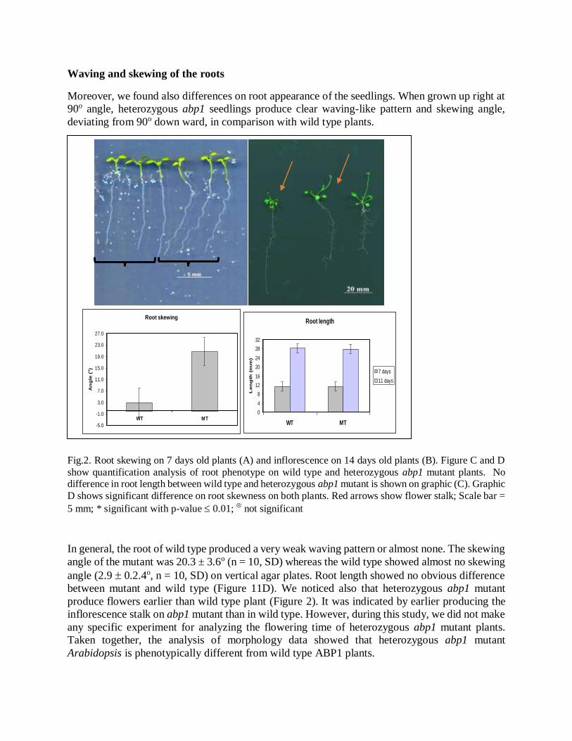
Waving and skewing of the roots
Moreover, we found also differences on root appearance of the seedlings. When grown up right at
90o angle, heterozygous abp1 seedlings produce clear waving-like pattern and skewing angle,
deviating from 90o down ward, in comparison with wild type plants.
Fig.2. Root skewing on 7 days old plants (A) and inflorescence on 14 days old plants (B). Figure C and D
show quantification analysis of root phenotype on wild type and heterozygous abp1 mutant plants. No difference in root length between wild type and heterozygous abp1 mutant is shown on graphic (C). Graphic
D shows significant difference on root skewness on both plants. Red arrows show flower stalk; Scale bar =
5 mm; * significant with p-value 0.01; not significant
In general, the root of wild type produced a very weak waving pattern or almost none. The skewing
angle of the mutant was 20.3 ± 3.6o (n = 10, SD) whereas the wild type showed almost no skewing
angle (2.9 0.2.4o, n = 10, SD) on vertical agar plates. Root length showed no obvious difference
between mutant and wild type (Figure 11D). We noticed also that heterozygous abp1 mutant
produce flowers earlier than wild type plant (Figure 2). It was indicated by earlier producing the
inflorescence stalk on abp1 mutant than in wild type. However, during this study, we did not make
any specific experiment for analyzing the flowering time of heterozygous abp1 mutant plants.
Taken together, the analysis of morphology data showed that heterozygous abp1 mutant
Arabidopsis is phenotypically different from wild type ABP1 plants.
Root skewing
-5.0
-1.0
3.0
7.0
11.0
15.0
19.0
23.0
27.0
WT MT
An
gle
(o)
Root length
0
4
8
12
16
20
24
28
32
WT MT
Len
gth
(m
m)
7 days
11 days

Evaluation of expression levels of auxin-induced early genes
From semi-quantitative data we found that a concentration 0.03μM of IAA seemed to give no
significant response in the expression auxin-induced genes. Therefore, we decided to use only 0.1
M IAA in auxin treatment in quantitative RT-PCR. For quantitative real time PCR experiments,
four auxin-induced early genes were tested and analyzed. All primers for PCR amplification were
obtained from Paponov et al. (2008). Quantifying the relative changes in gene expression was
calculated based on the 2-CT
method (Livak and Schmittgen, 2001). By normalizing cycle
threshold value (Ct) of gene target with Ct value of specific reference gene, generally
housekeeping genes, and Ct value of calibrator sample (ie. without treatment or control sample),
relative value expression of target genes can be generated. The common equation for CT is [(Ct
gene of interest – Ct reference gene) time x – (Ct gene of interest – Ct reference gene) time 0]. The 2-
CT
method is a convenient way to analyze the relative changes in gene expression from real time
quantitative PCR experiment. This method makes several assumptions, including that the
efficiency of the PCR is close to 1 and the PCR efficiency of the target gene is similar to the
internal control gene (Livak and Schmittgen, 2001). For investigating the physiological changes
in gene expression, the relative expression ratio is adequate for most purposes (Pfafft, 2001). In
this experiment, 18S rRNA gene was used as endogenous standard gene.
In this experiment, the expression of control samples (water treated samples) in wild type and
heterozygous abp1 samples are shown in figure 13. As shown on figure 13, the starting point of
expression of IAA2 gene in wild type was statistically significantly different from wild type
expression (p-value 0.01) in heterozygous abp1 samples, whereas the others gene were not
different. Interestingly, by comparing the expression of those genes with water treated samples
from microarray data, it was found that the expression of IAA2, IAA11, and IAA20 genes of wild
type control samples from quantitative RT PCR have a similar relative expression pattern in
microarray data. IAA2 was expressed about 5 times higher than IAA11, whereas IAA20 was very
low expressed. However, IAA13 expression differed from quantitative RT PCR data. The reason
remains unexplained.
As shown on Figure 3, at t = 0 min, the expression of three tested genes was higher in wild type
samples than in heterozygous abp1 samples. The IAA20 gene in heterozygous abp1 sample was
expressed higher than in wild type at t = 0 min. A t = 0 min the expression of IAA11 and IAA13
genes in wild type were 1.5 times fold higher than in heterozygous abp1 mutant samples (not
statistically significant), and the IAA2 gene in wild type was expressed 2.8 fold higher than on
heterozygous abp1 (statistically significant). After 30 min of auxin treatment, all genes were found
to be expressed higher in wild type samples than in heterozygous abp1 samples, especially the
IAA20 gene. The statistical significance for the 30 min values was clear. However, after 60 minute
of auxin treatment, only IAA11 and IAA13 expression was weakly higher (1.2 -1.7 fold) in wild
type samples in comparison to heterozygous abp1 samples. Those expression data indicate that
wild type plant expressed Aux/IAA genes (IAA3, IAA5, IAA14, and IAA20) higher than in
heterozygous abp1 mutant plants when treated by 0.1 M auxin.
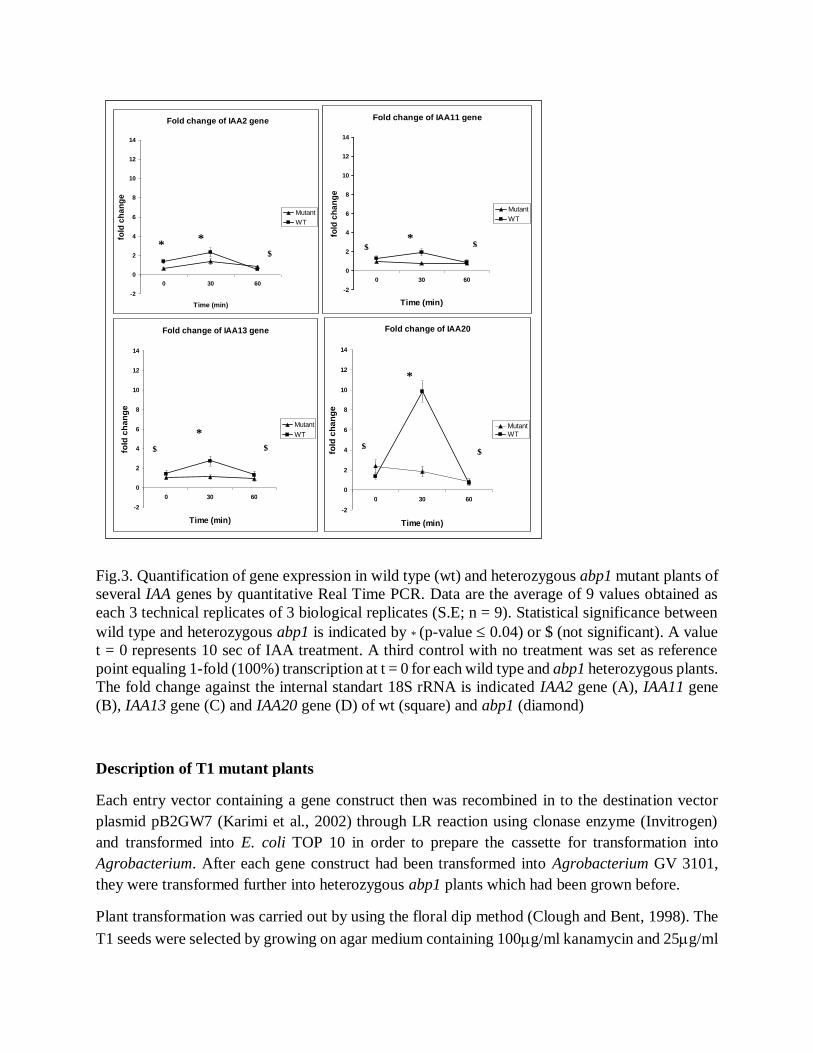
Fig.3. Quantification of gene expression in wild type (wt) and heterozygous abp1 mutant plants of
several IAA genes by quantitative Real Time PCR. Data are the average of 9 values obtained as
each 3 technical replicates of 3 biological replicates (S.E; n = 9). Statistical significance between
wild type and heterozygous abp1 is indicated by * (p-value 0.04) or $ (not significant). A value
t = 0 represents 10 sec of IAA treatment. A third control with no treatment was set as reference
point equaling 1-fold (100%) transcription at t = 0 for each wild type and abp1 heterozygous plants.
The fold change against the internal standart 18S rRNA is indicated IAA2 gene (A), IAA11 gene
(B), IAA13 gene (C) and IAA20 gene (D) of wt (square) and abp1 (diamond)
Description of T1 mutant plants
Each entry vector containing a gene construct then was recombined in to the destination vector
plasmid pB2GW7 (Karimi et al., 2002) through LR reaction using clonase enzyme (Invitrogen)
and transformed into E. coli TOP 10 in order to prepare the cassette for transformation into
Agrobacterium. After each gene construct had been transformed into Agrobacterium GV 3101,
they were transformed further into heterozygous abp1 plants which had been grown before.
Plant transformation was carried out by using the floral dip method (Clough and Bent, 1998). The
T1 seeds were selected by growing on agar medium containing 100g/ml kanamycin and 25g/ml
Fold change of IAA2 gene
-2
0
2
4
6
8
10
12
14
0 30 60
Time (min)
fold
ch
an
ge
Mutant
WT
Fold change of IAA11 gene
-2
0
2
4
6
8
10
12
14
0 30 60
Time (min)
fold
ch
an
ge
Mutant
WT
Fold change of IAA13 gene
-2
0
2
4
6
8
10
12
14
0 30 60
Time (min)
fold
ch
an
ge
Mutant
WT
Fold change of IAA20
-2
0
2
4
6
8
10
12
14
0 30 60
Time (min)
fold
ch
an
ge
MutantWT
* *
$ *
$
*
*
$ $ $
$ $
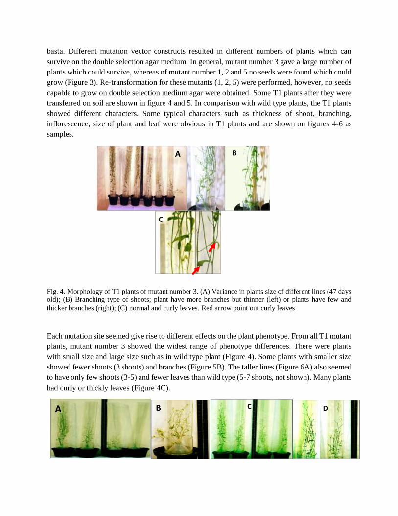
basta. Different mutation vector constructs resulted in different numbers of plants which can
survive on the double selection agar medium. In general, mutant number 3 gave a large number of
plants which could survive, whereas of mutant number 1, 2 and 5 no seeds were found which could
grow (Figure 3). Re-transformation for these mutants (1, 2, 5) were performed, however, no seeds
capable to grow on double selection medium agar were obtained. Some T1 plants after they were
transferred on soil are shown in figure 4 and 5. In comparison with wild type plants, the T1 plants
showed different characters. Some typical characters such as thickness of shoot, branching,
inflorescence, size of plant and leaf were obvious in T1 plants and are shown on figures 4-6 as
samples.
Fig. 4. Morphology of T1 plants of mutant number 3. (A) Variance in plants size of different lines (47 days old); (B) Branching type of shoots; plant have more branches but thinner (left) or plants have few and
thicker branches (right); (C) normal and curly leaves. Red arrow point out curly leaves
Each mutation site seemed give rise to different effects on the plant phenotype. From all T1 mutant
plants, mutant number 3 showed the widest range of phenotype differences. There were plants
with small size and large size such as in wild type plant (Figure 4). Some plants with smaller size
showed fewer shoots (3 shoots) and branches (Figure 5B). The taller lines (Figure 6A) also seemed
to have only few shoots (3-5) and fewer leaves than wild type (5-7 shoots, not shown). Many plants
had curly or thickly leaves (Figure 4C).
A B
C
A B C D
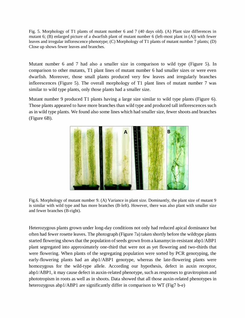
Fig. 5. Morphology of T1 plants of mutant number 6 and 7 (40 days old). (A) Plant size differences in
mutant 6; (B) enlarged picture of a dwarfish plant of mutant number 6 (left-most plant in (A)) with fewer leaves and irregular inflorescence phenotype; (C) Morphology of T1 plants of mutant number 7 plants; (D)
Close up shows fewer leaves and branches.
Mutant number 6 and 7 had also a smaller size in comparison to wild type (Figure 5). In
comparison to other mutants, T1 plant lines of mutant number 6 had smaller sizes or were even
dwarfish. Moreover, those small plants produced very few leaves and irregularly branches
inflorescences (Figure 5). The overall morphology of T1 plant lines of mutant number 7 was
similar to wild type plants, only those plants had a smaller size.
Mutant number 9 produced T1 plants having a large size similar to wild type plants (Figure 6).
Those plants appeared to have more branches than wild type and produced tall inflorescences such
as in wild type plants. We found also some lines which had smaller size, fewer shoots and branches
(Figure 6B).
Fig.6. Morphology of mutant number 9. (A) Variance in plant size. Dominantly, the plant size of mutant 9
is similar with wild type and has more branches (B-left). However, there was also plant with smaller size
and fewer branches (B-right).
Heterozygous plants grown under long-day conditions not only had reduced apical dominance but
often had fewer rosette leaves. The photograph (Figure 7a) taken shortly before the wildtype plants
started flowering shows that the population of seeds grown from a kanamycin-resistant abp1/ABP1
plant segregated into approximately one-third that were not as yet flowering and two-thirds that
were flowering. When plants of the segregating population were sorted by PCR genotyping, the
early-flowering plants had an abp1/ABP1 genotype, whereas the late-flowering plants were
homozygous for the wild-type allele. According our hypothesis, defect in auxin receptor,
abp1/ABP1, it may cause defect in auxin-related phenotype, such as responses to gravitropism and
phototropism in roots as well as in shoots. Data showed that all those auxin-related phenotypes in
heterozygous abp1/ABP1 are significantly differ in comparison to WT (Fig7 b-e)
A B
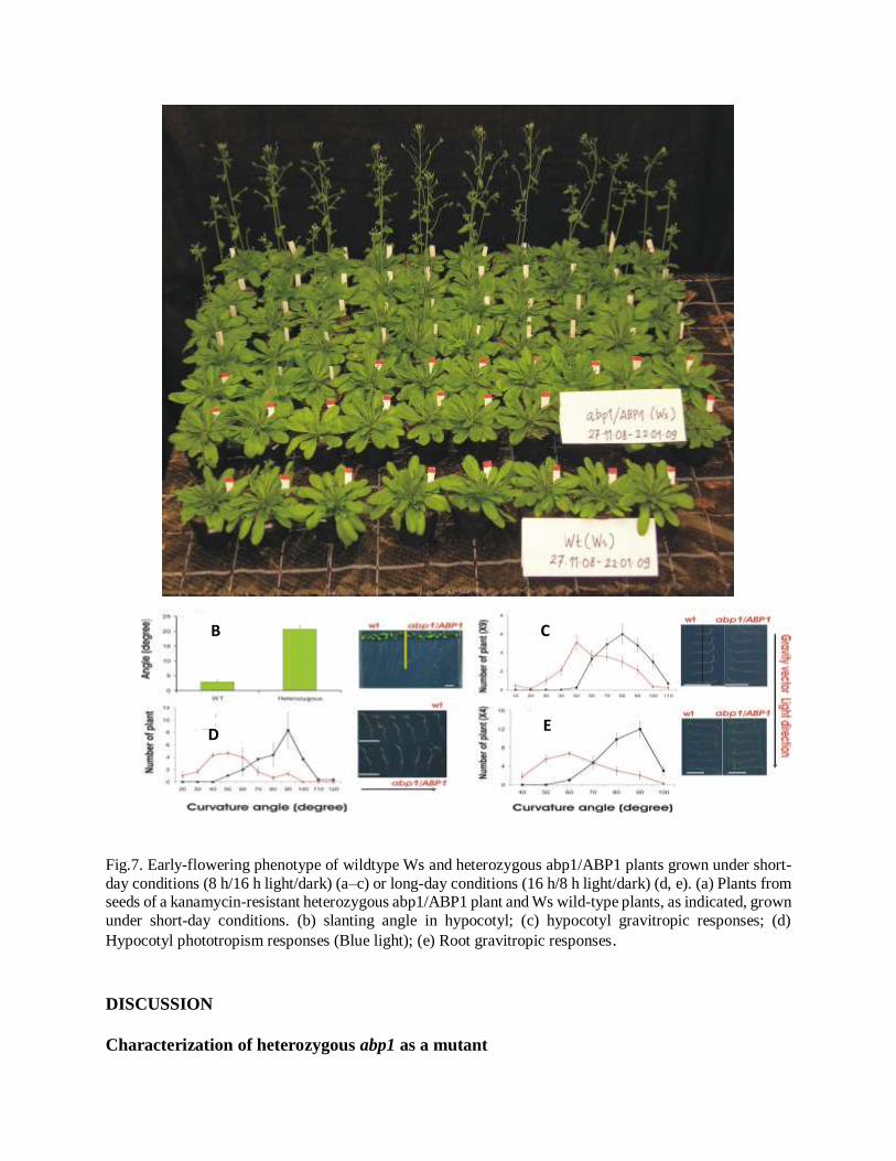
Fig.7. Early-flowering phenotype of wildtype Ws and heterozygous abp1/ABP1 plants grown under short-
day conditions (8 h/16 h light/dark) (a–c) or long-day conditions (16 h/8 h light/dark) (d, e). (a) Plants from seeds of a kanamycin-resistant heterozygous abp1/ABP1 plant and Ws wild-type plants, as indicated, grown
under short-day conditions. (b) slanting angle in hypocotyl; (c) hypocotyl gravitropic responses; (d)
Hypocotyl phototropism responses (Blue light); (e) Root gravitropic responses.
DISCUSSION
Characterization of heterozygous abp1 as a mutant
B C
D E
Before all gene constructs containing mutated ABP1 cDNA were transformed into heterozygous
abp1 plants, characterization the heterozygous abp1 mutant plants was performed. Our primary
observation was that heterozygous abp1 A. thaliana (Ws-2), in general, had similar characteristic
such as described by Chen et al. (2001). Analysis of the ABP1 gene in heterozygous abp1 plants
and wild type plant showed that heterozygous abp1 mutant plants produced a 1.5 kb fragment and
2.6 kb fragment in a PCR test technique, whereas using the same primers combination, wild type
plants produced only 2.6 kb fragment. This verified that the heterozygous abp1 mutant has two
different ABP1 alleles, one with and without insertion. The abp1 mutant seeds which were used in
this experiments and in Chen’s experiments originated from the same seed sources, both are from
Alan Jones group who donated abp1 mutant seeds to The Arabidopsis Biological Resource Center
– Ohio University (ABRC). Heterozygous abp1 mutant plants produced brown seed (wild type
and heterozygous abp1 seeds) and white seeds (homozygous abp1 seeds) in ratio 3:1 as predicted
by Mendel’s law in self mating of heterozygous organisms with one mutant allele. This is both
consistent with Chen et al. (2001).
However, further observations on seedlings and mature heterozygous abp1 T-DNA mutant plants
showed that the plants have a mutant morphological phenotype. Data were observed on seedling
stage, immature plants and mature plants have shown clearly differences between heterozygous
abp1 plants and wild type, which in Chen et al. (2001) did not mentioned. Interestingly, we found
that two white seeds could grow and produced a complete seedling and one mature green plant,
although after further growing for 3 weeks on soil this plant also died for unknown reasons.
According Chen et al. (2001), white seeds from heterozygous abp1 progenies cannot grow to
produce viable plant, because the embryo in all white seeds cannot develop to produce normal
embryo. Moreover, the second observation was that four weeks old heterozygous abp1 plants
showed clearly plants having a smaller size in comparison to wild type plants of equal age. Thirdly,
abp1 heterozygous plants also had a clear seedling phenotype showing a waving pattern and a
skewing angle when growing on vertical agar plate. The fourth observation was that heterozygous
abp1 plants flowered earlier than wild type although this was not quantified. Experiments for
evaluating the expression level of appropriate auxin-induced genes were also performed. Even
though, due to time restrictions, only few genes could be investigated and properly quantified by
qRT-PCR, clear differences in the expression of early auxin-induced genes could also be found in
the mutant. It is obvious that the heterozygous state of the ABP1 gene already is sufficient to make
the heterozygous abp1 plant a mutant. ABP1 very clearly is an excellent auxin binding protein
(Napier et al., 2002; Woo et al., 2002) so that it is difficult to assume something else but auxin
functions to be associated with it. The lack of chromatid expressing ABP1 mRNA may result in
lower mRNA and, subsequently, lower protein amounts. For reasons of the short time for this
project, mRNA of ABP1 was not tried to quantified but reduced amounts of ABP1 protein seem
to be most adequate explanation for the observed phenotype. If ABP1 is an auxin receptor this
should result in lower signal strength because of a lower number of active receptor molecules,
provided the same hormone concentration is kept. Lower signal strength should result in partial
losses of functions.
Can reduce steam size, early flowering, root waving, and altered regulation of auxin-induced genes
be interpreted as partial loss functions?. The reduced size of the abp1 heterozygous plants can
indeed be explained as a partial loss of elongation. Other mutants related to auxin signaling
showing reduced size are, for instance, axr1 (Lincoln et al., 1990), axr2-1 (Timpte, 1994), sax1
(Ephritikhine et al., 1999), acl5 (Imai et al., 2006), mya2 (Holweg and Nick, 2004), etc. Thus, in
this study, it is quite obvious that the reduced size phenotype is entirely consisted with a function
of ABP1 as an auxin receptor.
Early flowering was indicated by our data although it was not quantified. This rather points out to
some link to light signaling because Arabidopsis thaliana is a facultative long day plant.
Facultative long day plants flower earlier, for instance, if red light signaling by phytochrome B is
impeded (Fankhauser and Staiger, 2004). Other factors may also induce such a phenotype (Taiz
and Zeiger, 2006). It should be pointed out in this context, that activity of the ABP1 protein is
decreased by red light (Jones et al., 1991). This aspect seems worth investigating in further studies
on the heterozygous abp1 mutant.
The root waving phenotype of the young abp1 seedlings could also be related to auxin functions.
Waving mutant are associated with several gene groups most of which are related to a loss of
gravity sensing (Simmons et al., 1995; Simmons et al., 2006) and/or auxin transport defects
(agr1/eir1/pin2) or phototrophism (wav2) (Chen et al., 1999; Mochizukia et al., 2005). Other gene
defects leading to a waving mutant are also described which, presently, cannot be related to auxin
functions (Sedbrook et al., 1999). Lack of gravisensing or genetic defects can contribute to a
mutation in the waving pattern, circumnutation and negative thigmotropism an obstacle avoiding
response (Migliaccio and Piconese, 2001). Circumnutation also requires auxin transport and
gravisensing (Simmons et al., 1995) so that auxin transport and gravisensing disturbances are
major contributions to waving phenotypes, other gene defects cannot be excluded (Chen et al.,
1998). However, most waving mutants seem to be associated with auxin transport which is
integrated into gravitropism and phototropism. Recently, it was found that auxin itself inhibits
auxin transport by inhibiting the endocytosis of PIN protein (Paciorek et al., 2005). This inhibition
requires an auxin receptor other than TIR1 and several authors suggested this (Tomasz and Friml,
2006; Merks et al., 2007) and ABP1 could be a receptor for auxin inhibition of auxin efflux
transport which is faster regulated then gene regulation the only know function of TIR1 (Paciorek
et al., 2005; Calderon-Villalobos et al., 2006; Merks et al., 2007). So, recent publication point out
that there could be a link of ABP1 to PIN1 endocytosis recycling and auxin transport regulation
which might be a basis for a hypothesis stimulating further experiments on the waving phenotype
of abp1 heterozygous mutant. Such experiments could be investigation of gravitropic or
phototropic sensitivity or direct measurement of auxin transport.
It is obvious that the data suggest that ABP1 protein is an auxin receptor: abp1 heterozygous plants
have a phenotype affected in several auxin-regulated features, like stem elongation, root waving
and regulation of know auxin-regulated genes.
Analysis of auxin-induced early gene expression
Gene expression measured by quantitative real time PCR data.
Currently, there is no doubt that PCR can be made quantitative. In a number of publications, the
quantitative capability of PCR has been demonstrated by comparing it to classical means of nucleic
acids quantifying, such as Northern blot or Southern blot analysis (Ferre, 1992). Introduction of
quantitative real time PCR has shown that this new technique can be used to quantify more
precisely the amplification product. However, this technique require some condition in order to
get precision data, such as better primer performance (no dimer), precision on pipetting, the same
amount of DNA/cDNA template etc. Optimizing of the PCR reaction includes the cycle number
and right melting temperature also are essential conditions for this technique. Another important
factor in quantitative real time PCR experiments is the reference gene. Because the measurement
accuracy of gene expression in real time PCR generally relies on normalization of the expression
of target gene to specific reference gene, therefore the reference gene is a crucial factor for
obtaining a good result. Housekeeping genes are generally used as reference genes such as 18S
rRNA, UBIQUITIN (UBQ), ACTIN (ACT), b-TUBULIN (TUB), and glyceraldehyde-3-phosphate
dehydrogenase (GAPDH) because they were supposed to have a uniform expression (Jain et al.,
2006). Failure to use an appropriate control gene may result in biased gene expression profiles, as
well as low precision. The consequences may be that only gross changes in expression level are
declared statistically significant, or that patterns of expression are erroneously characterized
(Brunner et al., 2004).
We compared the expression of IAA2, IAA11, IAA13, and IAA20. Based on Ct values which have
been normalized by using Ct value of 18S rRNA gene, fold change of tested-genes were obtained.
Predominantly, the expression Aux/IAA genes in wild type samples differ significantly in
comparison with heterozygous abp1 samples, especially after 30 min auxin exposure (significant
with a p-value < 0.03). Only IAA2 did not show a significant difference after 30 min auxin
application. After zero time auxin exposure, the expression of IAA2 and IAA11 showed a
significant difference (significant with p-value 0.02) between wild type samples and
heterozygous abp1 samples. There are no statistically significant differences between wild type
samples and heterozygous abp1 samples on the expression of all tested genes in this experiment
at 60 min after auxin exposure. Those data show that all tested genes were varying their expression
at different times after auxin application and the changes were transient. Currently, there is no
information on whether ABP1 is involved in rapid transcriptional responses to auxin (Kepinski
and Leyser, 2005) but the data here clearly indicate this. However, several phenotypic differences
in wild type and heterozygous abp1 plants in this study and other publications about auxin-induced
cellular processes such as swelling of the protoplast (Steffens et al., 2001), stomatal closure (Blatt
and Thiel, 1994; Gehring et al., 1998), etc, indicated that it is probable any expression genes are
modulated via ABP1 perception.
Regarding differences of Aux/IAA genes expression data on wild type and heterozygous abp1
samples, there are some possibility reasons that those genes did not show clearly differences.
Several publications have reported that auxin induces the expression of many, but not all, Aux/IAA
gene family members. For example the Arabidopsis IAA1 through IAA14 and IAA19 genes are
auxin inducible with varying induction kinetics, and their mRNA accumulation varies in different
parts of the plant (Abel et al., 1995; Tatematsu et al., 2004). Several Aux/IAA genes, including
IAA17 and IAA28, show little or no response to exogenous auxin. The diversity in auxin
responsiveness and tissue-specific expression among the various gene family members suggests
that each member may have a distinct or overlapping function(s) during normal auxin responses
required for plant development (Overvoorde et al., 2005).
Paponov et al. (2008) demonstrated that a 0.5 h application of 1μM 1-NAA was enough to alter
IAA11 and IAA13 transcription. After 1 h exposure with 1μM 1-NAA both IAA11 and IAA13 genes
and also IAA2 gene elevated to a two fold change of transcription. However, IAA20 gene does not
show any alteration in transcription on the same condition. A high heterogeneity is observed in the
transcription of the Aux/IAA gene family in response to auxin in different microarray experiments
by using different auxins and plant material (Paponov et al., 2008). However, in comparison with
Paponov et al. (2008), there are two factors which differ compared to our experiment. First, we
used IAA instead of 1-NAA, and second, we used a very low IAA concentration 0.1μM for
induction gene expression, whereas Paponov et al. (2008) used 1μM 1-NAA. Furthermore, they
used Arabidopsis cell culture as plant material. It may be possibly that those factors give a
significant contribution to the differences. ABP1 has higher affinity to 1-NAA than to IAA (Löbler
and Klämbt, 1985; Yamagami et al., 2004). The affinity of ABP1 for 1-NAA is reported variously
as being between 50 and 200 nM, whereas its affinity for IAA is much lower between 5–10 mM
(Badescu and Napier, 2006) and it is affected by pH (Brown and Jones, 1994). The effect of auxin
also thought to depend on its concentration, with high and low doses eliciting different responses
(Teale et al, 2006). Nevertheless, the existing evidence suggests that there are multiple auxin
receptors, and hence the work on ABP1 is expected to answer only part of the question of how the
auxin signal is perceived.
Mutagenesis of some conserved amino acid residues of ABP1
Mutagenesis has been carried out in this project and all gene constructs have been successfully
obtained (supplement 1, 2). Nine gene constructs containing different mutations were transformed
into heterozygous abp1 mutant plants and T1 generation has been collected. Clear differences in
phenotypes of these T1 plants were observed and different mutations seemed to give different
effects on the phenotypes of the heterozygous abp1 transformed plants. Some characters which
have been collected concerning with T1 mutants are different size of plant, less branching, the
number of shoot, leaf appearance, etc (Figure 4-7, supplement 2).
The long objective of this experiment is actually to design clear homozygous abp1 mutant plants
which are not lethal so then further experiments with plants having mutant ABP1 protein can be
performed. Although selecting by using double marker (kanamycin and basta resistances) has been
carried out and T1 progenies containing both these gene markers were obtained, homozygous
ABP1 progeny was not yet obtained. By self mating, selecting using both markers, and PCR assays
of the further T generations (T2, T3, T4, T5), we believe that homozygous mutated-ABP1 T-DNA
insertion progenies with no expression of wild type ABP1 can be collected. However, at the
moment the work reached T1 selection for preparing T2 generations.
From T1 generation of different mutant lines, differences of plant phenotype were noticed.
Although we cannot prove that those phenotypic differences are due to mutation of given amino
acid residue(s), the reason for different phenotypes could be that the mutated protein is expressed
from the strong promoter whereas the wild type ABP1 is expressed from a weak wild type
promoter. Conceivably, mutant denies of ABP1 may dominate the phenotype. Currently, properly
experiments for testing this hypothesis cannot easily Currently, properly experiments for testing
this hypothesis cannot easily be performed, because the genetic background of those mutated-
ABP1 plants are not yet homozygous and the original wild type ABP1 protein remains in those
plants.
We also noticed on those T1 generations that some mutant lines are not able to grow on selective
medium (containing kanamycin and basta markers). Mutant lines contain mutations on tryptophan
135 (mutant number 1 and 2) and on tryptophan 151 (mutant number 5) showed those inability to
grow on selective agar medium. We noticed also that even though mutant number 4 and 6 which
contain mutation on tryptophan 135 were able to grow on selective medium, only few seedlings
could grow properly. Currently, we also encountered problems in growing mutant number 4 for
producing the next T generations. This could indicate that the mutant proteins are lethal to
seedlings.
Multi-alignment analysis showed that those amino acid residues used for mutation in this project
are all conserved. It suggests that those amino acids are likely to have important functions in ABP1
protein. All amino acid targets for mutagenesis this experiment actually have been described in
previous publications in which those amino acid residues were predicted to be involved in the
binding of auxin by ABP1 protein or in the folding of ABP1 protein. Warwickers et al. (2001) in
their publication proposed that there are two tripeptide motifs on ABP1, DDW136 and WDE153,
which either one of these tripeptides could occupy the auxin-binding site in the absence of ligand.
Displacement of this tripeptide by free auxin would induce a conformation change to initiate
signaling. Similar result was also reported by David et al. (2001) working with Nicotiana
tabaccum. They concluded that site-directed mutagenesis on WDE175, corresponding to WDE153
on Arabidopsis, have a critical role in protein folding and functional activity of ABP1 at the plasma
membrane. Moreover, Woo et al. (2002) described that the binding pocket of ABP1 is
predominantly hydrophobic with a metal ion deep inside the pocket coordinated by three histidines
and a glutamate. Auxin binds within this pocket with its carboxylate group binding the zinc and
its aromatic ring binding hydrophobic residues including Trp151. All together suggests that
tryptophan 135 and 151 have crucial functions in ABP1 protein. According to crystal study of
ABP1 protein of maize, Woo et al. (2002) also revealed that Cys155 has a crucial function on
ABP1 protein. Together with Cys2, Cys155 form a single disulfide bridge which important for
stabilizing the protein both in the presence and absence of bound auxin. No mutation of cysteines
was attempted.
However, much works remain need to be done in order to get a better understanding of the function
of amino acid residue(s) of ABP1. By obtaining clear homozygous mutated ABP1 plant lines, we
expect that better experiments to study ABP1 functions can be performed. Nevertheless, currently
we have proven that introduction of new mutated ABP1 gene constructs into Arabidopsis was
successfully carried out. For future works, the next T generations will be collected in order to
obtaining homozygous abp1 mutant plants.
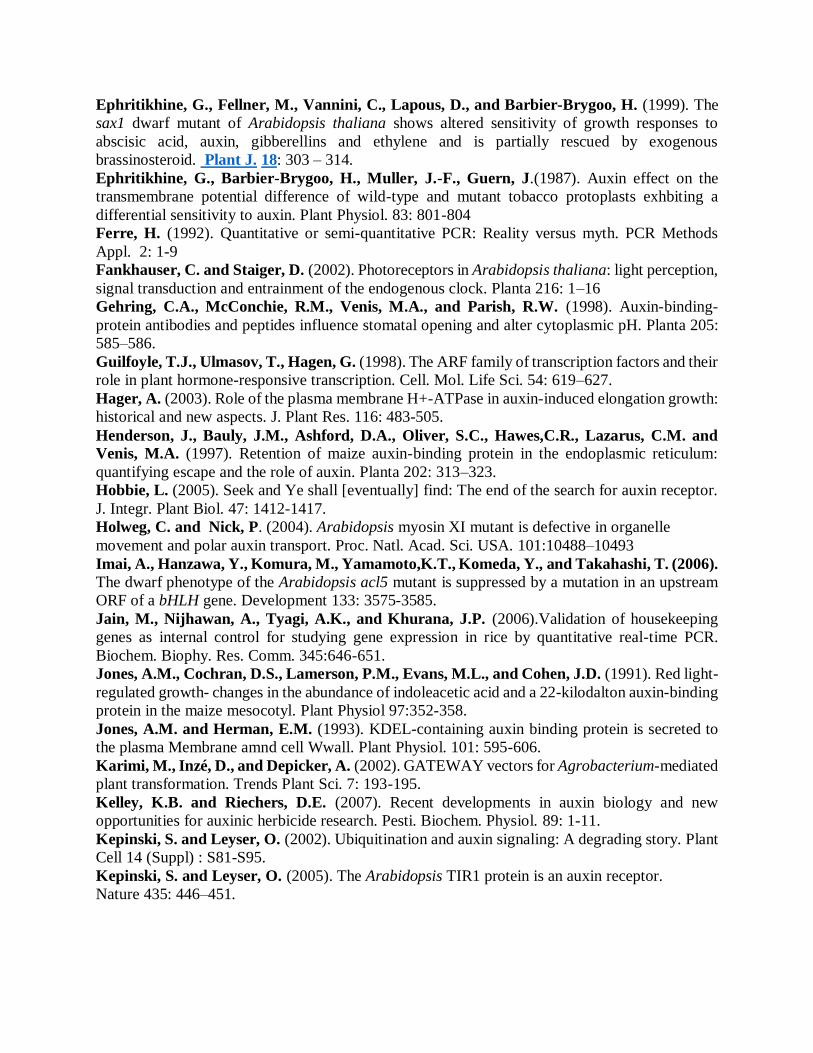
REFERENCES
Abel, S., Nguyen, M.D., and Theologis, A. (1995). The PS-IAA4/5-like family of early auxin-
inducible mRNAs in Arabidopsis thaliana. J. Mol. Biol. 251: 533–549
Abel, S. and Theologis, A. (1996). Early genes and auxin action. Plant Physiol. 111: 9-11
Azpiroz-Leehan R, Feldmann KA. (1997).T-DNA insertion mutagenesis in Arabidopsis: going
back and forth. Trends Genet. 113:152-156.
Badescu, G.O., and Napier, R.M. (2006). Receptors for auxin: will it all end in TIRs?. Trends
Plant Sci. 11: 217-223.
Barbier-Brygoo H, Ephritikhine G, Klambt D, Ghislain, M., and Guem J. (1989). Functional
evidence for an auxin receptor at the plasmalemma of tobacco mesophyll protoplasts. Proc. Natl.
Acad. Sci, USA 86: 891-895.
Bauly, JM, Sealy, IM, Macdonald, H, Brearley, J, Droge, S, Hillmer, S, Robinson, DG, Venis,
MA, Blatt, MR, Lazarus, CM & Napier, RM. (2000). Overexpression of auxin-binding protein
enhances the sensitivity of guard cells to auxin. Plant Physiol. 124:1229 - 1238.

Berleth, T., and Sachs, T. (2001). Plant morphogenesis: Long-distance coordination and local
patterning. Curr. Opin. Plant Biol. 4: 57–62.
Blatt, M.R. and Thiel, G. (1994). K+ channels of stomatal guard cells: bimodal control of the
inward-rectifier evoked by auxin. Plant J. 5: 55-68.
Brown, J.C. and Jones, A.M. (1994). Mapping the auxin-binding site of auxin-binding protein 1.
J. Biol. Chem. 269: 21136–21140
Brunner, A.M., Yakovlev, I.A., and Strauss, S.H. (2004). Validating internal controls for
quantitative plant gene expression studies. BMC Plant Biol. 4:14.
Calderon-Villalobos, L.I.A., Kuhnle, C., Li, H., Rosso, M., Weisshaar, B., and
Schwechheimer, C. (2006). LucTrap vectors are tools to generate luciferase fusions for the
quantification of transcript and protein abundance in vivo1. Plant Physiol.141: 3–14.
Campanoni, P. and Nick, P. (2005). Auxin-dependent cell division and cell elongation 1-
Naphthaleneacetic acid and 2,4-dichlorophenoxyacetic acid activate different pathways. Plant
Physiol. 137: 939–948
Chen, J.G., Ullah, H., Young, J.C., Sussman, M.R. and Jones, A.M. (2001). ABP1 is required
for organized cell elongation and division in Arabidopsis embryogenesis. Genes Dev. 15: 902–911
Chen, R., Hilson, P., Sedbrook, J., Rosen, E., Caspar, T. and Masson, P.H. (1998). The
Arabidopsis thaliana AGRAVITROPIC1 gene encodes a component of the polar auxin-transport
efflux carrier. Proc. Natl. Acad. Sci. U. S. A. 95: 15112–15117.
Chen, R., Rosen, E., and Masson, P.H. (1999). Gravitropism in higher plants. Plant Physiol.
120: 343-350
Chen, Y.F., Randlett, M.D., Findell, J.L., and Schaller. G.E. (2002). Localization of the
ethylene receptor ETR1 to the endoplasmic reticulum of Arabidopsis. J. Biol. Chem. 277: 19861-
19866.
Claussen, M., Lüthen, H., Blatt, M., and Böttger, M. (1997). Auxin-induced growth and its
linkage to potassium channels. Planta 201:227–234.
Clough, S.J. and Bent, A.F. (1998). Floral dip: a simplified method for Agrobacterium-mediated
transformation of Arabidopsis thaliana. Plant J. 16: 735–743.
David, K., Carnero-Diaz, E., Leblanc, N., Monestiez, M., Grosclaude, J. and Perrot-
Rechenmann, C. (2001). Conformational dynamics underlie the Activity of the auxin-binding
protein, Nt-abp1. J. Biol. Chem. 276: 34517-34523.
Delker,C., Raschke, A., Quint, M. (2008). Auxin dynamics: the dazzling complexity of a small
molecule’s Message. Planta.227:929–941
Deineko, E.V.,Zagorskaya, A.A.,and Shumny, V.K. (2007). T-DNA-induced mutations in
transgenic plants. Russian J. Gen. 43: 1–11.
Dharmasiri, N., Dharmasiri, S. and Estelle, M. (2005). The F-box protein TIR1 is an auxin
receptor. Nature 435: 441-445.
Dharmasiri, N. and Estelle, M. (2004). Auxin signaling and regulated protein degradation.
Trends Plant Sci. 9: 302–308
Dharmasiri, N., Dharmasiri, S., Jones, A. M. and Estelle, M. (2003). Auxin action in a cell
free system. Curr. Biol. 13: 1418-1422.
Diekmann W, Venis MA, Robinson DG. (1995). Auxin induces clustering of the auxin-binding
protein at the surface of division in Arabidopsis embryogenesis. Genes Dev. 15: 902–911
Edgerton, M.D., Tropsha, A. and Jones, A.M. (1994). Modelling the auxin-binding site of
auxin-binding protein1 of maize. Phytochem. 35: 1111–1123.
Ephritikhine, G., Fellner, M., Vannini, C., Lapous, D., and Barbier-Brygoo, H. (1999). The
sax1 dwarf mutant of Arabidopsis thaliana shows altered sensitivity of growth responses to
abscisic acid, auxin, gibberellins and ethylene and is partially rescued by exogenous
brassinosteroid. Plant J. 18: 303 – 314.
Ephritikhine, G., Barbier-Brygoo, H., Muller, J.-F., Guern, J.(1987). Auxin effect on the
transmembrane potential difference of wild-type and mutant tobacco protoplasts exhbiting a
differential sensitivity to auxin. Plant Physiol. 83: 801-804
Ferre, H. (1992). Quantitative or semi-quantitative PCR: Reality versus myth. PCR Methods
Appl. 2: 1-9
Fankhauser, C. and Staiger, D. (2002). Photoreceptors in Arabidopsis thaliana: light perception,
signal transduction and entrainment of the endogenous clock. Planta 216: 1–16
Gehring, C.A., McConchie, R.M., Venis, M.A., and Parish, R.W. (1998). Auxin-binding-
protein antibodies and peptides influence stomatal opening and alter cytoplasmic pH. Planta 205:
585–586.
Guilfoyle, T.J., Ulmasov, T., Hagen, G. (1998). The ARF family of transcription factors and their
role in plant hormone-responsive transcription. Cell. Mol. Life Sci. 54: 619–627.
Hager, A. (2003). Role of the plasma membrane H+-ATPase in auxin-induced elongation growth:
historical and new aspects. J. Plant Res. 116: 483-505.
Henderson, J., Bauly, J.M., Ashford, D.A., Oliver, S.C., Hawes,C.R., Lazarus, C.M. and
Venis, M.A. (1997). Retention of maize auxin-binding protein in the endoplasmic reticulum:
quantifying escape and the role of auxin. Planta 202: 313–323.
Hobbie, L. (2005). Seek and Ye shall [eventually] find: The end of the search for auxin receptor.
J. Integr. Plant Biol. 47: 1412-1417.
Holweg, C. and Nick, P. (2004). Arabidopsis myosin XI mutant is defective in organelle
movement and polar auxin transport. Proc. Natl. Acad. Sci. USA. 101:10488–10493
Imai, A., Hanzawa, Y., Komura, M., Yamamoto,K.T., Komeda, Y., and Takahashi, T. (2006).
The dwarf phenotype of the Arabidopsis acl5 mutant is suppressed by a mutation in an upstream
ORF of a bHLH gene. Development 133: 3575-3585.
Jain, M., Nijhawan, A., Tyagi, A.K., and Khurana, J.P. (2006).Validation of housekeeping
genes as internal control for studying gene expression in rice by quantitative real-time PCR.
Biochem. Biophy. Res. Comm. 345:646-651.
Jones, A.M., Cochran, D.S., Lamerson, P.M., Evans, M.L., and Cohen, J.D. (1991). Red light-
regulated growth- changes in the abundance of indoleacetic acid and a 22-kilodalton auxin-binding
protein in the maize mesocotyl. Plant Physiol 97:352-358.
Jones, A.M. and Herman, E.M. (1993). KDEL-containing auxin binding protein is secreted to
the plasma Membrane amnd cell Wwall. Plant Physiol. 101: 595-606.
Karimi, M., Inzé, D., and Depicker, A. (2002). GATEWAY vectors for Agrobacterium-mediated
plant transformation. Trends Plant Sci. 7: 193-195.
Kelley, K.B. and Riechers, D.E. (2007). Recent developments in auxin biology and new
opportunities for auxinic herbicide research. Pesti. Biochem. Physiol. 89: 1-11.
Kepinski, S. and Leyser, O. (2002). Ubiquitination and auxin signaling: A degrading story. Plant
Cell 14 (Suppl) : S81-S95.
Kepinski, S. and Leyser, O. (2005). The Arabidopsis TIR1 protein is an auxin receptor.
Nature 435: 446–451.
Kim, Y.S., Min, J.K., Kim, D. and Jung, J. (2000). A soluble auxin binding protein, ABP57.
Purification with anti-bovine serum albumin antibody and characterization of its mechanistic role
in the auxin effect on plant plasma membrane H+-ATPase. J. Biol. Chem. 276: 10730–10736.
Klämbt, D. (1990). A views about the function of auxin binding proteins at the plasma membrane.
Plant Mol. Biol. 14: 1045–1050.
Krysan, P.J., Young, J.C., and Sussman, M.R. (1999). T-DNA as an insertional mutagen in
Arabidopsis. Plant Cell. 11: 2283–2290
Leblanc, N., David, K., Grosclaude, J., Pradier, J. M., Barbier Brygoo, H., Labiau, S., and
Perrot-Rechenmann, C. (1999). A novel immunological approach establishes that the auxin-
binding protein, Ntabp1, is an element involved in auxin signaling at the plasma membrane. J.
Biol. Chem. 274: 28314-28320.
Leyser, O. (2001). Auxin signaling: The beginning, the middle and the end. Curr. Opin. Plant
Biol. 4: 382-386.
Leyser, O. (2002). Molecular genetic of auxin signaling. Annu. Rev. Plant Biol. 53: 377-398.
Lincoln, C., Britton, J.H., and Estelle, M. (1990). Growth and development of the axr1
mutants of Arabidopsis. Plant Cell 2: 1071–1080.
Livak, K.J. and Schmittgen, T.D. (2001). Analysis of Relative Gene Expression Data Using
Real-Time Quantitative PCR and the 2-CT Method. Methods 25: 402–408.
Löbler, M. and Klämbt, D. (1985). Auxin-binding protein from coleoptile membranes of corn
(Zea mays L.). I. Purification by immunological methods and characterization. J. Biol. Chem.
260: 9848–9853.
Macdonald H. (1997). Auxin perception and signal transduction. Physiol. Plant.100: 423–430.
Merks, R.M.H., Van de Peer, Y., Inze, D., Beemster, G.T.S. (2007). Canalization without flux
sensors: a traveling-wave hypothesis. Trends Plant Sci. 12:384-390.
Migliaccio, F. and Piconese, S. (2001). Spiralizations and tropisms in Arabidopsis roots. Trends
Plant Sci. 6:561–565.
Mochizukia, S., Haradaa, A., Inadaa, S., Sugimoto-Shirasub, K., Staceyb, N., Wadac, T.,
Ishiguroc, S., Okadaa, K., and Sakai, T. (2005). The Arabidopsis WAVY GROWTH 2 protein
modulates root bending in response to environmental stimuli. Plant Cell 17:537-547.
Murashige,T and Skoog, F. (1962). A revised medium for rapid growth and bioassays with
tobacco tissue cultures. Physiol. Plant 15: 473-497.
Napier RM. (2001). Models of auxin binding. J. Plant Growth Regul. 20: 244–254.
Napier, R.M., David, K.M., and Perrot-Rechenmann, C. (2002). A short history of auxin-
binding proteins. Plant Mol. Biol. 49: 339–348
Napier, R.M., Fowke, L.C., Hawes, C., Lewis, M., and Pelhara H.R.B. (1992). Immunological
evidence that plants use both HDEL and KDEL for targeting proteins to the endoplasmic
reticulum. J. Cell Sci. 102: 261-71.
Oono, Y., Ooura,C., and Uchimiya, H. (2002). Expression pattern of Aux/IAA genes in the
iaa3/shy2-1D mutant of Arabidopsis thaliana (L.). Ann. Bot. 89: 77-82.
Ostergaard, L. and Yanofsky, M.F. (2004). Establishing gene function by mutagenesis in
Arabidopsis thaliana. Plant J. 39: 682-696.
Overvoorde, P.J., Okushima, Y., Alonso, J.M. Chan, A., Chang, C., Ecker, J.R., Hughes, B.,
Liu, A., Onodera, C., Quach, H., Smith, A., Yu, G., and Theologis, A. (2005). Functional
genomic analysis of the AUXIN/INDOLE-3-ACETIC ACID gene family members in Arabidopsis
thaliana. Plant Cell 17: 3282–3300.
Paciorek, T., Zazímalová, E., Ruthardt, N., Petrásek, J., Stierhof, Y.D., Kleine-Vehn, J.,
Morris, D.A., Emans, N., Jürgens, G., and Geldner, N. (2005). Auxin inhibits endocytosis and
promotes its own efflux from cells. Nature 435: 1251–1256
Paponov, I,A., Paponov, M., Tealea, W., Mengesb, M., Chakraborteeb, S., Murray, J.A.H.,
and Palmea, K. (2008). Comprehensive transcriptome analysis of auxin responses in
Arabidopsis. Mol. Plant 1: 321–337.
Parry, G. and Estelle, M. (2006). Auxin receptors: a new role for F-box proteins. Curr. Opin.
Cell Biol. 18: 152-156.
Pennazio, S. (2002). The discovery of the chemical nature of the plant hormone auxin. Rev. Biol.
95: 289–308.
Pfaffl, M.W. (2001). A new mathematical model for relative quantification in real-time RT-PCR.
Nucleic Acids Res. 29:e45.
Rück, A., Palme, K., Venis, M. A., Napier, R. M. and Felle, H. (1993). Patch-clamp analysis
establishes a role for an auxin binding protein in the auxin stimulation of plasma membrane current
in Zea mays protoplasts. Plant J. 4: 41-46.
Rügger, M., Dewey, E., Gray, W.M., Hobbie, L., Turner, J., and Estelle, M. (1998). The TIR1
protein of Arabidopsis functions in auxin response and is related to human SKP2 and yeast Grr1p.
Genes and Dev. 12: 198-207.
Schwechheimer, C. and Villalobos, L.I.A. (2004). Cullin-containing E3 ubiquitin ligases in plant
development. Curr. Opin. Plant Biol. 7: 677 – 686.
Sedbrook, J.C., Chen, R., and Masson, P.H. (1999). ARG1 (Altered Response to Gravity)
encodes a DnaJ-like protein that potentially interacts with the cytoskeleton. Proc. Natl. Acad.
Sci. USA. 96:1140–1145.
Simmons, C., Migliaccio, F., Masson, P., Caspar, T. and Soll, D. (1995). A novel root
gravitropism mutant of Arabidopsis thaliana exhibiting altered auxin physiology. Physiol. Plant.
93: 790–798.
Simmons, C., Migliaccio, F, Masson, P., Caspar, T., and Söll, D. (2006). A novel root
gravitropism mutant of Arabidopsis thaliana exhibiting altered auxin physiology. Physiol Planta.
93:790 – 798.
Steffens, B., Feckler, C., Palme, K., Christian, M., Böttger, M. and Lüthen, H. (2001). The
auxin signal for protoplast swelling is perceived by extracellular ABP1. Plant J. 27: 591–999.
Sussman, M.R., Amasino, R.M., Young, J.C., Krysan, P.J., and Phillips, S.A. (2000).The
Arabidopsis knockout facility at the University of Wisconsin–Madison1. Plant Physiol. 124: 1465-
1467.
Taiz,L. and Zeiger, E. (2006). Plant Physiology. 4th Ed. Palgrave Macmillan
Tan, X., Calderon-Villalobos, L.I.A., Sharon, M., Zheng, C., Robinson, C.V., Estelle, M., and
Zheng, N. (2007). Mechanism of auxin perception by the TIR1 ubiquitin ligase. Nature 446: 640-
645.
Tatematsu, K., Kumagai, S., Muto, H., Sato, A., Watahiki, M.K., Harper, R.M., Liscum, E.,
and Yamamoto, K.T. (2004). MASSUGU2 encodes Aux/IAA19, an auxin-regulated protein that
functions together with the transcriptional activator NPH4/ARF7 to regulate differential growth
responses of hypocotyl and formation of lateral roots in Arabidopsis thaliana. Plant Cell 16: 379–
393.
Teale, W.D., Paponov, I.A, and Palme, K. (2006). Auxin in action: signalling, transport and the
control of plant growth and development. Nat. Rev. Mol. Cell Biol. 7: 847-859.

Thiel, G., Blatt, M.R., Fricker, M.D., White, I.R., and Millner, P. (1993). Modulation of K+
channels in Vicia stomatal guard cells by peptide homologs to the auxin binding protein C
terminus. Proc. Natl. Acad. Sci. USA. 24: 11493-11497.
Tian, H., Klämbt, D., and Jones, A.M. (1995). Auxin binding protein 1 does not bind auxin
within the endoplasmic reticulum despite this being the predominant sub-cellular location for this
hormone receptor. J. Biol.Chem. 270: 26962–26969.
Timpte, C., Wilson, A.K., and Estelle, M. (1994). The axr2-1 Mutation of Arabidopsis thaliana
is a gain-of-function mutation that disrupts an early step in auxin response. Genetics 138: 1239-
1249.
Tomasz, P. and Friml, J. (2006). Auxin signalling. J. Cell Sci. 119: 1199-1202.
Venis, M.A. and Napier, R.M. (1995). Auxin receptors and auxin binding proteins. Crit. Rev.
Plant Sci. 14: 27–47.
Warwicker, J. (2001). Modelling of auxin-binding protein 1 suggests that its C-terminus and
auxin could compete for a binding site that incorporates a metal ion and tryptophan residue 44.
Planta 212: 343–347.
Woo, E.J., Marshall, J., Bauly, J., Chen, J.G., Venis, M., Napier,R. and Pickersgill, R. (2002).
Crystal structure of auxin-binding protein 1 in complex with auxin. EMBO J. 21: 2877–2885.
Woodward, A.W. and Bartel, B. (2005). A Receptor for auxin. Plant Cell 17: 2425-2429.
Yamagami, M., Haga, K., Napier, R.M., Iino, M., Leblanc, N., Roux, C., Pradier, J.M. and
Rechenmann, P. (2004). A two distinct signaling pathways participate in auxin-induced swelling
of pea epidermal protoplasts. Plant Physiol. 134: 735–747.
Zeiger, E. (2000). Sensory transduction of blue light in guard cells. Trends Plant Sci. 5: 183–185.
Zheng, L., Baumann, U., and Reymond, J.L. (2004). An efficient one-step site-directed and site-
saturation mutagenesis protocol. Nucl. Acids Res. 32:e115.
Supplement 1 – Multiple sequence alignment of ABP1 gene in mutants
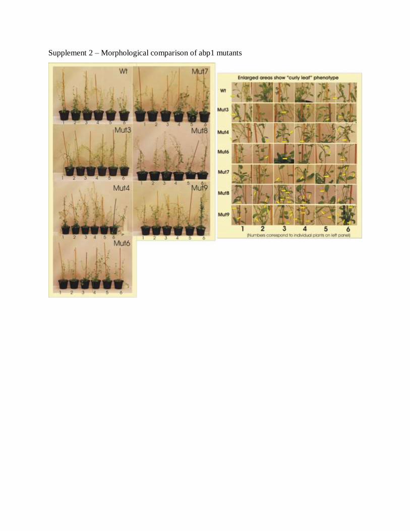
Supplement 2 – Morphological comparison of abp1 mutants