radiofrecuencia para reduccion de medidas
DESCRIPTION
El articulo describe el uso de una nueva tecnologia, la radiofrecuencia selectiva para reduccion de la grasa coroporalTRANSCRIPT
Lasers in Surgery and Medicine 45:235–239 (2013)
Operator Independent Focused High Frequency ISM Bandfor Fat Reduction: Porcine Model
Robert Weiss, MD, FAAD, FACPh,*1 Margaret Weiss, MD, FAAD,1 Karen Beasley, MD, FAAD,1
Jan Vrba, PhD,2 and Jan Bernardy, PhD2
1MD Laser, Skin & Vein Institute, Baltimore, Maryland2Czech Technical University, Prague, Czech Republic
Background: Selective fat reduction has been clearlyshown for various methods and energy modalities includ-ing cryolipolysis and high intensity focused thermalultrasound. Mathematical modeling of focused highfrequency of the EM spectrum has indicated that selectiveheating of fat is possible using wavelengths not previousexplored. The purpose of this study was to demonstrate inthe porcine model that selective heating of fat is possiblewith a non-contact, operator independent device.Methods: High frequencies of the Industrial, Scientificand Medical (ISM) RF band were utilized to reduceabdominal fat in a porcine model. Practical application ofmathematical modeling allowed an auto-feedback loop tobe developed to allow operator independent adjustment ofenergy to maintain subcutaneous fat at 45–468C whileoverlying skin remained at 40–418C.Results: Treatments of three Vietnamese pigs wereperformed under anesthesia in a certified veterinaryfacility. Gross and microscopic histologic results demon-strated a marked reduction in adipocytes of the treatedarea after 4 treatments of a total of 30 minutes each, withincremental fat diminution after each treatment. A final70% reduction of the abdominal fat layer was seen in thetreated areas. Duplex ultrasound revealed a reduction offat layer from 7.6 to 2.9 mm. Histologic evaluationrevealed that epidermis, dermis, and adnexal structuressuch as hair follicles were unaffected by the treatment,while adipocytes were significantly affected.Conclusion: A new model of fat reduction using highfrequency RF has been successfully achieved in a porcinemodel. This has very positive implications in the develop-ment of an operator independent, contact free devicefor reduction of fat in clinical practice. Lasers Surg. Med.45:235–239, 2013. � 2013 Wiley Periodicals, Inc.
INTRODUCTION
Multiple modalities to induce adipocyte apoptosis inorder to reduce pockets of fat non-invasively have recentlybecome available. These modalities primarily aim attargeting the properties of fat which differentiate itfrom skin and muscle, thus resulting in selective removalor dissolution of fat otherwise known as lipolysis.Currently available non-invasive fat removal methodsutilize heating or cooling utilizing, laser, radiofrequency,
and ultrasound sources to more selectively target adipo-cytes [1–5].
Themedical use of RF is based on an oscillating electricalcurrent forcing collisions between charged molecules andions which are then transformed into heat. A dielectric,such as fat, is an insulator with the ability of innerpolarization. Adipose tissue contains electrical dipoles.The direction of dipoles is chaotic and polarizationarranges dipoles in one direction. Dielectric polarizationrequires that every electrical dipole is rotated against thepolarization of the electrical field. With a rapidly alternat-ing electromagnetic field, all electrical dipoles oscillate.This oscillating movement of dipoles leads to heating up ofdipoles of fatty tissue, a principle mechanism of action ofhigh frequency on fat.
This study was designed to investigate an operatorindependent focused field system designed for contactlessdeep tissue thermal energy application. The applicator–generator circuitry is engineered to selectively deliver theenergy to tissue layer with specific impedance, in this casethe adipose tissue layer. This high frequency systemfocuses energy specifically into the adipose tissue layer,while limiting the delivery to the dermis, epidermis andmuscles. A multipolar broad field applicator shapes theelectro-magnetic field to optimize the penetration andmaximize the treatment area. Using a patented EnergyFlowControl (EFC) system this device automatically tunesthe tissue-applicator–generator circuitry to selectivelydeliver the energy to the adipose tissue layer whileminimizing the risk of overheating of the skin, muscles,or internal organs as is detailed below.
Conflicts of Interest Disclosures: BTL Aesthetics, Prague, CRprovided funding for this study. Dr. Robert Weiss is a speaker forBTL. The MD Laser, Skin & Vein Institute received equipmentfrom BTL.
This study was accepted for presentation in abstract form forthe 2013 ASLMS Annual Conference, Boston, MA.�Correspondence to: Robert A. Weiss, MD, FAAD, FACPh,Associate Professor, Johns Hopkins U School of Medicine;Director, MD Laser, Skin & Vein Institute, 54 Scott Adam Road,Hunt Valley, Baltimore, MD 21030.E-mail: [email protected] www.mdlsv.com www.smoothskin.net
Accepted 13 March 2013Published online in Wiley Online Library(wileyonlinelibrary.com).DOI 10.1002/lsm.22134
� 2013 Wiley Periodicals, Inc.
METHODS
This study was approved by the Institutional AnimalCare and Use Committee (IACUC) and the Committee forAnimal Protection of the Ministry of Agriculture of theCzech Republic. Procedures used conformed to acceptedpractices and to minimize or avoid causing pain, distress,or discomfort to the animals. In those circumstances inwhich study procedures were likely to cause more thanmomentary or slight pain or distress, the animals receivedanalgesics or anesthetics as per the Institutional AnimalCare and Use Committee (IACUC) at University ofVeterinary and Pharmaceutical Sciences Brno.
Four square shaped areas of the skin in rectusabdominalis (each square 10 cm � 10 cm) were selectedand marked as A, B, C, and D. Hairs were removed fromthe application area by clipping. The device used toperform treatment was the high frequency field RF devicesold as Vanquish, BTL Aesthetics, Prague, CR. Treatmentwas performed with the RF applicator placed approxi-mately 1 cm above the skin. Total exposure time (30 mi-nutes) of each treatment was divided into two 15 minutehalves. Skin surface was kept in the temperature rangefrom 39 to 428C during the treatment period.
Animals were under the total anesthesia and under thesupervision of the veterinarian during each treatment andduring the biopsy. Supervising veterinarians selected thetype and the dose of the anesthesia.
All animals were observed for clinical signs, morbidity ormortality once a day during the treatment period. Onset,duration and severity of any signs were recorded. Clinicalobservations included: (1) skin, eyes, and mucous mem-branes changes, (2) respiratory, circulatory, autonomic,and the central nervous system, (3) somatomotor activityand behavior pattern, changes in gait, posture andresponse to handling, presence of clonic or tonic move-ments and stereotypes.
Blood samples for clinical chemistry were collected fromall animals before each treatment, after each treatmentand 3 months after the last treatment (recovery period).Blood samples were collected into Tapval tubes withoutanticoagulation (clinical chemistry), serum samples wereobtained by centrifugation at 3,000 rpm for 15 minutes.Serum for clinical chemistry (�1 ml) was transferred intoappropriately labeled and sealed Eppendorf tube andfrozen at �208C or below until transport for analysis.
The treated area was examined before 1st, 3rd, 4thtreatment and at the end of the study period by Duplexultrasound. All animals were individually weighed beforeeach treatment and after the study period. Temperaturesof the epidermis and the adipose tissue temperature weremeasured by thermocouple before treatment, after 5, 12,15, 20, 27, and 30 minutes of the treatment and immedi-ately 30 minutes of exposure time. Epidermal temperaturewas monitored with an infrared camera during eachtreatment. Biopsy samples of the skin and the fat tissuewere taken with biopsy needle before treatment, after thefirst treatment, after 2nd, 4th treatment and 3 monthsafter the last treatment (recovery period). Samples of liver,
lung, and skin from all three swine for histopathologyexam were taken at the conclusion of the treatmentprotocol.
RESULTS
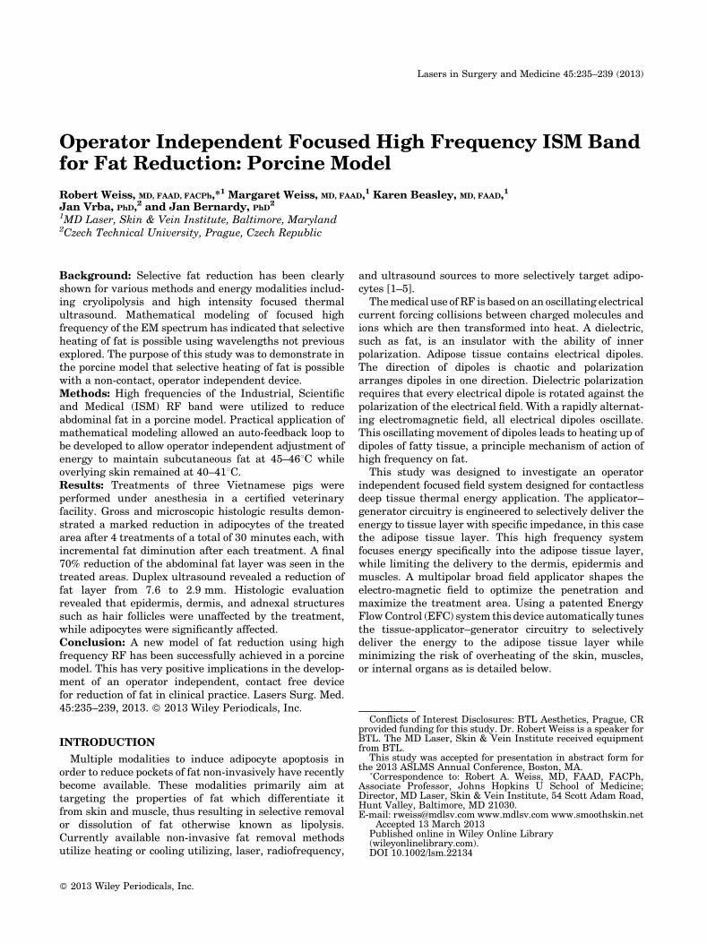
Skin erythema was observed during treatment, usuallyseveral minutes after the start of the treatment. Skinerythema resolved within minutes following the cessationof treatment. The first biopsy revealed infiltration of foamymacrophages and neutrophil granulocytes (Fig. 1).
Fig. 1. A: Normal fat before treatment.B: Disrupted fat after 4thtreatment. C: Foamy macrophages following treatment.
236 WEISS ET AL.
Biopsies taken 2 weeks after the third treatment werewithout signs of panniculitis. Histological analysis of skinbiopsy samples taken after first treatment revealeddesquamation of superficial layers of epidermis, perivas-cular infiltration, fragmentation of superficial collagenfibrils, and alteration of the adipose tissue. Significantadipose tissue destruction was found (Fig. 1).Following histological examinations after the second and
fourth treatment showed similar signs as observed afterthe first treatment with desquamation of superficial layersof epidermis, perivascular infiltration, fragmentation ofsuperficial collagen fibrils, and alteration of the adiposetissue with the exception of sporadic coagulo-necroticlesions of the adipose tissue. After the fourth treatment thehistological evaluation showed adipose tissue destructionin dermal layers. In one subject more frequent fibroticsepta were found after the fourth treatment.Histological examination of the skin biopsy after the end
of the recovery period revealed in all animals localdesquamation of epidermal surface, sporadic perivascularinfiltration in dermis, focal disintegration in adiposetissue, and thick fibrotic septa.Histology of the skin samples from dermal depression
showed intense desquamation of epidermis, focal mononu-clear infiltration of dermal vessels, fragmentation ofcollagen fibrils, multicystic foci with inflammatory cellinfiltration. Adipose tissue was observed to be disruptedalong with focal fibrosis.The TUNEL method was used to detect apoptotic nuclei
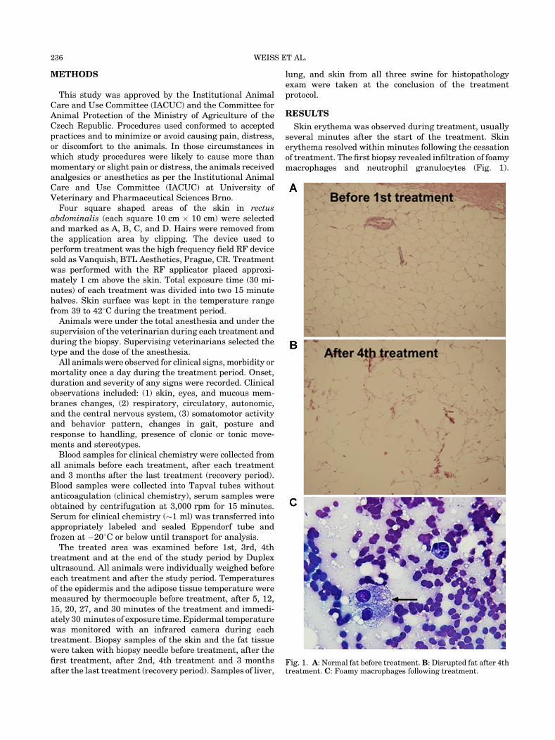
in the samples. Terminal deoxynucleotidyl transferasedUTP nick end labeling (TUNEL) is amethod for detectingDNA fragmentation by labeling the terminal end of nucleicacids. TUNEL is a common method for detecting DNAfragmentation that results from apoptotic signalingcascades. The assay relies on the presence of nicks in theDNA which can be identified by terminal deoxynucleotidyltransferase or TdT. It may also label cells that havesuffered severeDNAdamage. The detected apoptotic indexwas 13/100 prior to the initial treatment and 52/100following the last treatment. Evidence of apoptosis isshown in Figure 2.Serum levels of glucose, bilirubin, urea, ALT, total
cholesterol, HDL and LDL cholesterols and triglycerideswere unaffected by treatment. Aspartate aminotransfer-ase (AST) serum activity increase was observed in onesubject at one time point but was judged not related totreatment.
A final 70% reduction of the abdominal fat layerwas seenin the treated areas. Average adipose layer thicknessreduction was 6.9 mm. Duplex ultrasound revealed areduction of fat layer from 7.6 to 2.9 mm. No significantchanges in body weight were measured as individual bodyweights of the animals were within normal range,corresponding to the age and baseline weight.
Fig. 2. Evidence of apoptosis by TUNEL staining method.Fig. 3. Evolution of temperature over time from 0 to 15 minutes.A: Subject 1. B: Subject 2. C: Subject 3.
FOCUSED FIELD RF FOR FAT REDUCTION 237
The skin temperature and the adipose tissue tempera-ture measurement results were recorded in a separategraph for each animal subject. Figure 3A–C shows the firstpart of therapy (first 15 minutes). Room temperature was�228C. Before the therapy the deep tissues temperaturewas�358C. After 5 minutes of the treatment the tempera-
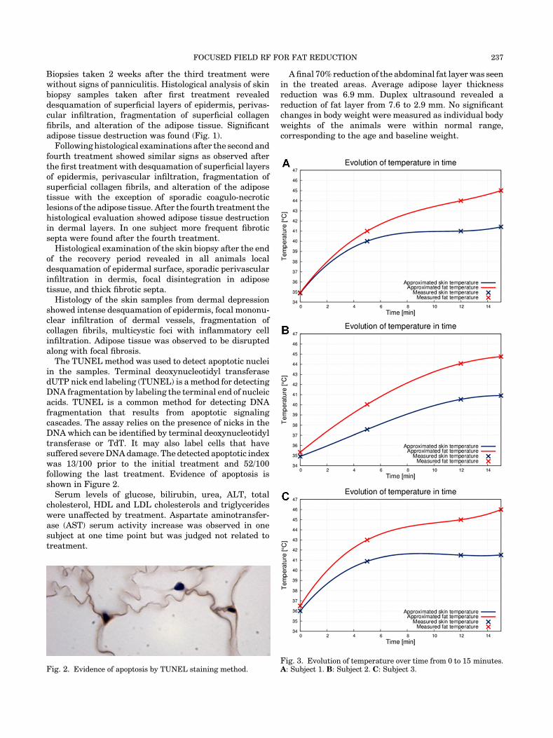
ture of the skin increased to 408C and the temperature ofthe adipose tissue increased to 428C. The effect of the EFCsystem contributing the selective heating of the adiposetissue layer is noticeable after 10 minutes of therapy.Temperature difference between the skin and the adiposetissue layer was 48C; skin temperature was 418C, and theadipose tissue layer temperature is 458C. Skin tempera-ture remained on the tolerable level while the adiposetissue temperature reached the desired therapeutic level.The second group of graphs shows the second half of thetherapy (next 15 minutes) (Fig. 4A–C). The broad electro-magnetic field heated whole treatment area. The tempera-ture of the adipose tissue layer increased approximately upto 44–458C.The device is engineered to primarily focus on the
adipose tissue layerwith specific impedance. This principleleads to the adipose tissue layer heating-up faster thanother tissue layers. Also blood-rich tissues layers such asskin and muscle are cooled down by the circulating bloodmuch faster than the adipose tissue layer. Furthermore,
Fig. 4. Evolution of temperature over time from 15 to 30 minutes.A: Subject 1. B: Subject 2. C: Subject 3.
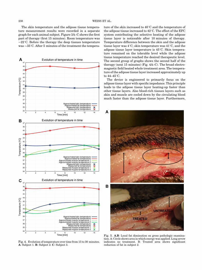
Fig. 5. A,B: Local fat diminution on gross pathologic examina-tion. A:Circle showsarea inwhich energywas applied. Long arrowindicates no treatment. B: Treated area shows significantreduction of fat in subject 2.
238 WEISS ET AL.
due to no contact application of the treatment head, theskin can be additionally cooled by circulating air.Gross pathology examination revealed local skin depres-
sions in all three animals. Skin section led through thelesion showed signs of an apoptotic reaction causingreduction of the adipose tissue layer (Fig. 5A,B). No othermacroscopic signs were observed during gross pathologyexamination.Duplex ultrasound examination was performed to
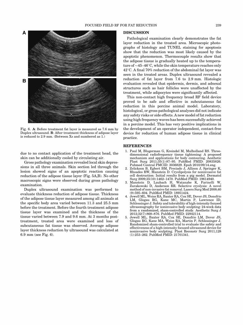
evaluate thickness reduction of adipose tissue. Thicknessof the adipose tissue layer measured among all animals atthe specific body area varied between 11.3 and 25.5 mmbefore the treatment. Before the fourth treatment adiposetissue layer was examined and the thickness of thetissue varied between 7.9 and 9.6 mm. At 3 months post-treatment, treated area were examined and loss ofsubcutaneous fat tissue was observed. Average adiposelayer thickness reduction by ultrasound was calculated at6.9 mm (see Fig. 6).
DISCUSSION
Pathological examination clearly demonstrates the fatlayer reduction in the treated area. Microscopic photo-graphs of histology and TUNEL staining for apoptosisshow that the reduction was most likely caused by theapoptotic phenomenon. Thermocouple results show thatthe adipose tissue is gradually heated up to the tempera-ture of�45–468C, while the skin temperature reaches only428C. A final 70% reduction of the abdominal fat layer wasseen in the treated areas. Duplex ultrasound revealed areduction of fat layer from 7.6 to 2.9 mm. Histologicevaluation revealed that epidermis, dermis, and adnexalstructures such as hair follicles were unaffected by thetreatment, while adipocytes were significantly affected.
This non-contact high frequency broad RF field deviceproved to be safe and effective in subcutaneous fatreduction in this porcine animal model. Laboratory,histological, or gross pathological analyses did not indicateany safety risks or side effects. A newmodel of fat reductionusing high frequencywaves has been successfully achievedin a porcine model. This has very positive implications inthe development of an operator independent, contact-freedevice for reduction of human adipose tissue in clinicalpractice.
REFERENCES
1. Paul M, Blugerman G, Kreindel M, Mulholland RS. Three-dimensional radiofrequency tissue tightening: A proposedmechanism and applications for body contouring. AestheticPlast Surg 2011;35(1):87–95. PubMed PMID: 20835826.Pubmed Central PMCID: 3036829. Epub 2010/09/14.eng.
2. Zelickson B, Egbert BM, Preciado J, Allison J, Springer K,Rhoades RW, Manstein D. Cryolipolysis for noninvasive fatcell destruction: Initial results from a pig model. DermatolSurg 2009;35(10):1462–1470. PubMed PMID: 19614940.
3. Manstein D, Laubach H, Watanabe K, Farinelli W,Zurakowski D, Anderson RR. Selective cryolysis: A novelmethod of non-invasive fat removal. Lasers SurgMed 2008;40(9):595–604. PubMed PMID: 18951424.
4. Jewell ML, Weiss RA, Baxter RA, Cox SE, Dover JS, DonofrioLM, Glogau RG, Kane MC, Martin P, Lawrence ID,Schlessinger J. Safety and tolerability of high-intensity focusedultrasonography for noninvasive body sculpting: 24-week datafrom a randomized, sham-controlled study. Aesthetic Surg J2012;32(7):868–876. PubMed PMID: 22942114.
5. Jewell ML, Baxter RA, Cox SE, Donofrio LM, Dover JS,Glogau RG, Kane MA, Weiss RA, Martin P, Schlessinger J.Randomized sham-controlled trial to evaluate the safety andeffectiveness of a high-intensity focused ultrasound device fornoninvasive body sculpting. Plast Reconstr Surg 2011;128(1):253–262. PubMed PMID: 21701341.
Fig. 6. A: Before treatment fat layer is measured as 7.6 mm byDuplex ultrasound. B: After treatment thickness of adipose layeris reduced to 2.9 mm. (Between Xs and numbered as 1.)
FOCUSED FIELD RF FOR FAT REDUCTION 239