daftar pustaka -...
TRANSCRIPT
51
DAFTAR PUSTAKA
1. Kementrian Kesehatan RI. Pusat data dan informasi kementrian kesehatan RI 2014.
Jakarta: Kementrian Kesehatan RI ; 2014.
2. Supraptono B. Korelasi antara penurunan sensibilitas kornea dengan retinopati
diabetika pada penderita diabetes mellitus. Semarang: Fakultas Kedokteran Universitas
Diponegoro; 1999.
3. Suyono S, Pandelaki K. Buku ajar ilmu penyakit dalam. Setiati S, Alwi I, Sudoyo A,
Simadibrata M, Setyohadi B, Syam AR, editor. Jakarta: InternaPublishing; 2014.
4. McCulloch DK. Diabetic retinopathy : pathogenesis. Netherland: Wolters Kluwer;
2015.
5. Riordan-Eva P, Wither JP. Vaughan & Asbury’s general ophthalmology. New York:
McGrawHill; 2004.
6. Heinemann B, House L, Hill J, Ox O. Eye essential. Philadelphia: Elsevier; 2008.
7. Riordan-Eva P. Vaughan & Asbury oftalmologi umum. Jakarta: Penerbit Buku
Kedokteran EGC; 2012.
8. American Optometric Association. Dry Eye. American Optometric Association. 2015
[updated 2011 Oct 15; cited 2015 Nov 17]. Available from
http://www.aoa.org/patients-and-public/eye-and-vision-problems/glossary-of-eye-and-
vision-conditions/dry-eye
9. Foster CS. Dry eye syndrome. Medscape. 2015 [updated 2012 Des 14; cited 2015 Des
20]. Available from http://emedicine.medscape.com/article/1210417
10. Figueroa-Ortiz LC, Jiménez Rodríguez E, García-Ben A, García-Campos J. Study of
52
tear function and the conjunctival surface in diabetic patients. Arch la Soc Española
Oftalmol. 2011; 86(4): 107-112.
11. Achtsidis V, Eleftheriadou I, Kozanidou E, et al. Dry eye syndrome in subjects with
diabetes and association with neuropathy. Diabetes care. 2014; 37: 210-211
12. Rahman A, Yahya K, Ahmed T, Sharif-Ul-Hasan K. Diagnostic Value of Tear Film Test in
type 2 diabetes. JMPA. 2007;57:577.
13. Manaviat, Masoud Reza Rashidi M, Afkhami-Ardekani M, Reza Shoja M. Prevalence of dry
eye syndrome and diabetic retinopathy in type 2 diabetic patients. BMC Opthamol. 2008;8..
14. Fong DS, Aiello L, Gardner TW, et al. Retinopathy in diabetes. Diabetes care. 2004;
27(1): 84-87.
15. Brownlee M. The pathobiology of diabetic complications a unifying mechanism.
Diabetes. 2005; 54(6):1615-1625.
16. Chew E. Diabetes mellitus a fundamental and clinical text. Philadelphia: Lippincolt
William and Willkins; 2000.
17. Fraser CE, D’Amico DJ. Diabetic retinopaty : classification and clinical features.
Netherlands: Wolters Kluwer; 2015.
18. Ding J, Sullivan DA. Aging and dry eye disease. Exp Gerontol. 2012;47(7):483-490.
19. Singh S, Moksha L, Sharma N, Titiyal JS, Biswas NR, Velpandian T. Development
and evaluation of animal models for sex steroid deficient dry eye. J Pharmacol Toxicol
Methods. 2014;70(1):29-34.
20. Ousler GW, Wilcox KA, Gupta G, Abelson MB, Fink K. An evaluation of the ocular
drying effects of 2 systemic antihistamines: loratadine and cetirizine hydrochloride.
53
Ann Allergy, Asthma Immunol. 2004;93(5):460-464.
21. Hitner H, Nagle B. Pharmacology : An Introcduction. New York: Mc Graw Hill; 2012.
22. Bowlinh E. Which oral meds cause dry eye. 2011[updated 2012 Nov 14; cited 2015
Des 20]. Available from http://www.reviewofcontactlenses.com/content/d/dry_eye
23. Fujita M, Igarashi T, Kurai T, Sakane M, Yoshino S, Takahashi H. Correlation
between dry eye and rheumatoid arthritis activity. Am J Ophthalmol. 2005;140(5):808-
813.
24. Karabulut GO, Kaynak P, Altan C, et al. Corneal biomechanical properties in thyroid
eye disease. Kaohsiung J Med Sci. 2014;30(6):299-304.
25. Lee JH, Kee CW. The Significance of tear film break up time in the diagnosis of dry
eye syndrome. Kor. J Ophtalmol. 1988;2:69-71.
26. Sweeney DF, Millar TJ, Raju SR. Tear film stability: a review. Exp Eye Res.
2013;117:28-38.
27. Lan W, Lin L, Xiao Y, Yu M. Automatic noninvasive tear break up time (TBUT) and
conventional fluorescent TBUT. Optom Vis Sci. 2014;91(12):1412-1418.
28. Murphy PJ. Noninvasive assessment of corneal sensitivity in young and elderly
diabetic and nondiabetic subjects. Invest Ophthalmol Vis Sci. 2004;45(6):1737-1742.
29.` Kim JH, Kim JH, Nam WH, et al. Oral alcohol administration disturbs tear film and
ocular surface. Ophthalmology. 2012;119(5):965-971.
30. Mengher LS, Pandher KS, Bron AJ. Non-invasive tear film break-uptime: sensitivity
and specificity. Acta Ophthalmol. 1986;64(4):441-444.
54
31. Massoud Reza Manaviat, Maryam Rashidi, Mohammad Afkhami-Ardekani,
Mohammad Reza Shoja. Prevalence of dry eye syndrome and diabetic retinopathy in
type 2 diabetic patients. BMC Opthalmology.2008;8:10
32. Hasan IN, Anggarwal P, Gurav A, Patel N. Assessment of dry eye status in type 2
diabetic patients in tertiary health care hospital, India. IOSR J Dent Med Sci.
2014;13(8):06-11.
33. Chibis GW. Fundamentals and Principles of Ophthalmology, Basic & Clinical Science
Course: Leo Publisher
34. Kaiserman I, Kaiserman N, Nakar S, Vinker S; Dry eye in diabetic patients; Am J
Ophthalmol. 2005 Mar; 139 (3): 498-503.
35. Kahn HA, Bradley RF. Prevalence of diabetic retinopathy Age, sex, and duration of
diabetes. Brit F Opthal. 1975;59(345).
36. Lee AJ, Lee J, Saw S-M, et al. Prevalence and risk factors associated with dry eye
symptoms: a population based study in Indonesia. Br J Ophtamol. 2002;86:1347-
1351.
37. Gilbar JP. Human tear film electrolyte concentrations in health and dry-eye disease.
int OpthamolClin. 1994;(3427):36.
38. Tanushree V, HV Gowda. Corelation of dry eye status with severity of diabetic
retinopathy. Retin J Evol Med Dent Sci. 2014;3(66):14323-14329
39. Kaur K. Menopause Hormone Replacement Therapy. Medscape.
http://emedicine.medscape.com/article/276104-overview#a6. Published 2016.
40. M Ozdemir,H Temizdemir. Age and gender related tear function changes in normal
population. Eye. 2010;24:79-83
55
41. Dogru M, Katamami C, Inoue M. Tear function and ocular surface changes in
noninsulin-dependent diabetes mellitus. Ophtalmology. 2001;108:586-592.
42. Citirik M, Berker N, Haksever H, Elgin U, Ustun H. Conjunctival impression cytology
in non-proliferative and proliferative diabetic retinopathy. Int J Ophthamol. 2014;7(2).
43. Najafi L,Malek M, Valojerdi AE,Aghili E,Khamseh ME,Fallah AE. Dry eye and its
correlation to diabetes microvascular complications in people with type 2 diabetes
mellitus. J Diabetes Complicat. 2013;27(5):459-462.
56
Lampiran 1. Informed consent
JUDUL PENELITIAN : Perbedaan Tear Film Break Up Time pada Pasien Retinopati
Diabetika Nonproliferatif Dibandingkan Retinopati Diabetika
Proliferatif
INSTANSI PELAKSANA : Bagian Mata FK Undip – Mahasiswa Program Studi Strata -1
Kedokteran Umum Fakultas Kedoteran Universitas
Diponegoro
PERSETUJUAN SETELAH PENJELASAN
(INFORMED CONSENT)
Berikut ini naskah yang akan dibacakan pada responden / ibu responden penelitian:
Yth. Bapak/Ibu/Sdr : ............................
Nama saya Michelle Abigail, mahasiswa Program Studi S1 Ilmu Pendidikan Dokter
Fakultas Kedokteran UNDIP. Saya akan melakukan penelitian dengan judul Perbedaan Tear
film break up time pada Pasien Retinopati Diabetika Nonproloferatif Dibandingkan Pasien
Retinopati Diabetika Proliferatif. Tujuan dari penelitian ini adalah untuk mencari perbedaan
tear film break up time pada pasien retinopati diabetika nonproliferatif dan pasien retinopati
diabetika proliferatif di RSUP Dr. Kariadi Semarang. Anda terpilih sebagai peserta penelitian
ini. Apabila Bapak/Ibu/Saudara setuju sebagai peserta penelitian maka akan ada beberapa hal
yang akan Bapak/IbuSaudara alami, yaitu :
1. Diminta beberapa informasi tentang data diri
2. Pemeriksaan tear film break up time dengan menggunakan fluoroscein strip dan
slitlamp
Pemeriksaan tear film break up time dilakukan dengan cara sebagai berikut :
1. Meletakkan secarik kertas fluoroscein yang sedikit dilembabkan pada
konjunctiva(mata) pasien, lalu meminta pasien untuk berkedip
2. Lapisan air mata kemudian diperiksa dengan bantuan filter cobalt pada slitlamp,
pasien diminta agar tidak berkedip
3. Mencatat waktu munculnya bintik kering pertama pada lapisan fluoroscein kornea
Tidak ada efek samping yang berbahaya pada pemeriksaan ini. Efek samping yang
mungkin terjadi adalah terdapat sisa-sisa zat fluoroscein pada mata, dimana efek samping
tersebut dapat ditangani. Penanganan pada sisa-sisa zat fluoroscein yaitu dibersihkan dengan
menggunakan akuades(air bersih) yang dialirkan pada mata.
57
Keuntungan bagi Bapak/Ibu/Saudara yang bersangkutan ikut dalam penelitian ini
adalah dapat mengetahui tear film break up time dimana hasil tersebut menjadi patokan
terjadinya sindroma mata kering yang sering terjadi pada penderita retinopati diabetika.
Saya menjamin bahwa setiap data pemeriksa dan penelitian akan dijamin kerahasiaanya, dan
data hanya akan saya gunakan untuk kepentingan penelitian. Sebagai peserta penelitian
keikutsertaan ini bersifat sukarela dan tidak dikenakan biaya penelitian. Apabila ada
informasi yang belum jelas atau pertanyaan mengenai penelitian ini Bapak/Ibu/Saudara bisa
menghubungi saya Michelle Abigail, manasiswi Program Studi S1 Ilmu Pendidikan Dokter
FK UNDIP (085747182653)
Terima kasih atas kerjasama Bapak/Ibu/Saudara
Sudah mendengar dan memahami penjelasan penelitian, dengan ini saya menyatakan
SETUJU / TIDAK SETUJU
untuk ikut sebagai subyek/sampel penelitian ini.
Semarang, …………………… 2016.
Saksi 1 :
Nama Terang : Nama Terang :
Alamat : Alamat :
Saksi 2 :
Nama Terang :
Alamat :
58
PERBEDAAN TEAR FILM BREAK UP TIME PADA PASIEN RETINOPATI
DIABETIKA NONPROLIFERATIF DIBANDINGKAN DENGAN RETINOPATI
DIABETIKA PROLIFERATIF
Nama :.................................................................................................
Usia :.................................................................................................
Jenis kelamin : Laki-laki / Perempuan*
Pendidikan :.................................................................................................
Pekerjaan :.................................................................................................
Alamat :.................................................................................................
Lama Menderita DM : .................................................................................................
59
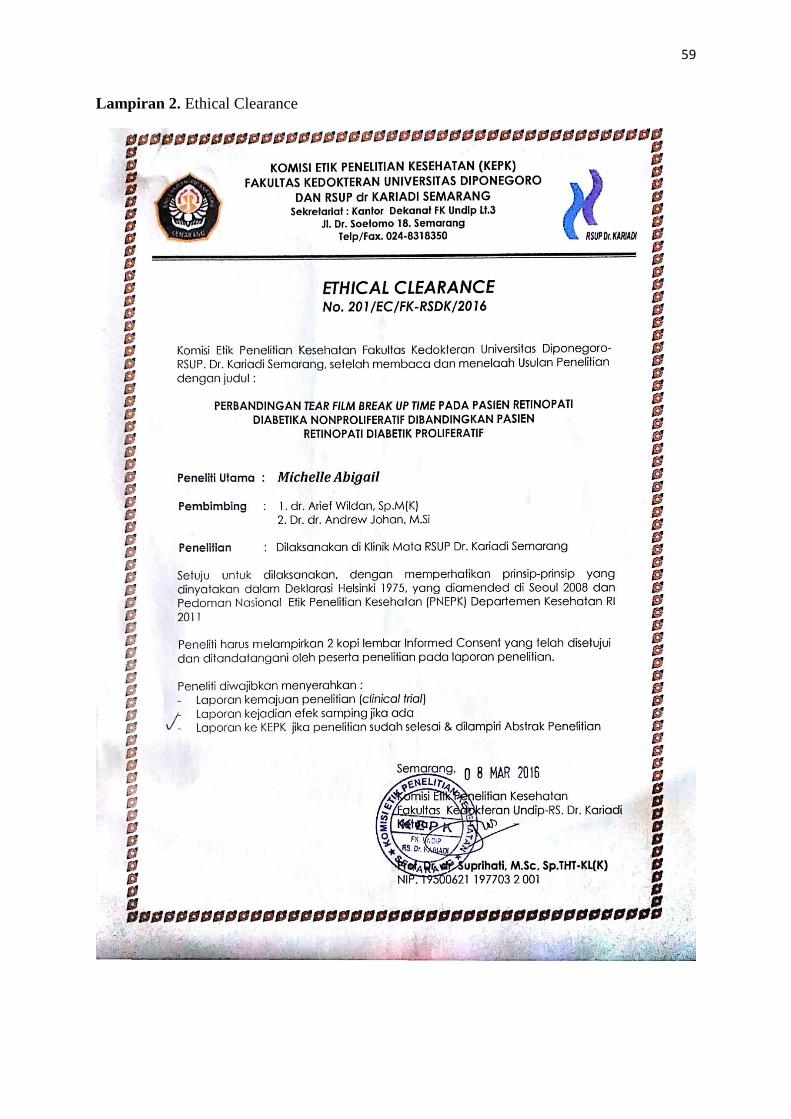
Lampiran 2. Ethical Clearance
60
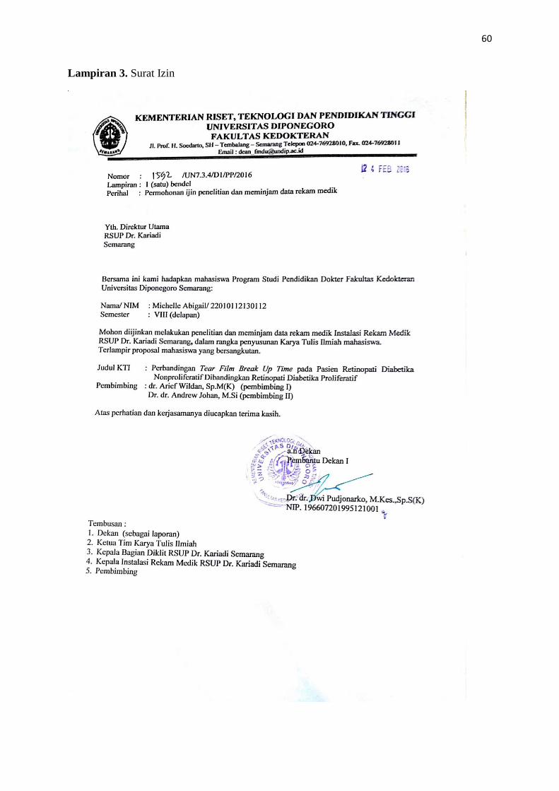
Lampiran 3. Surat Izin
61
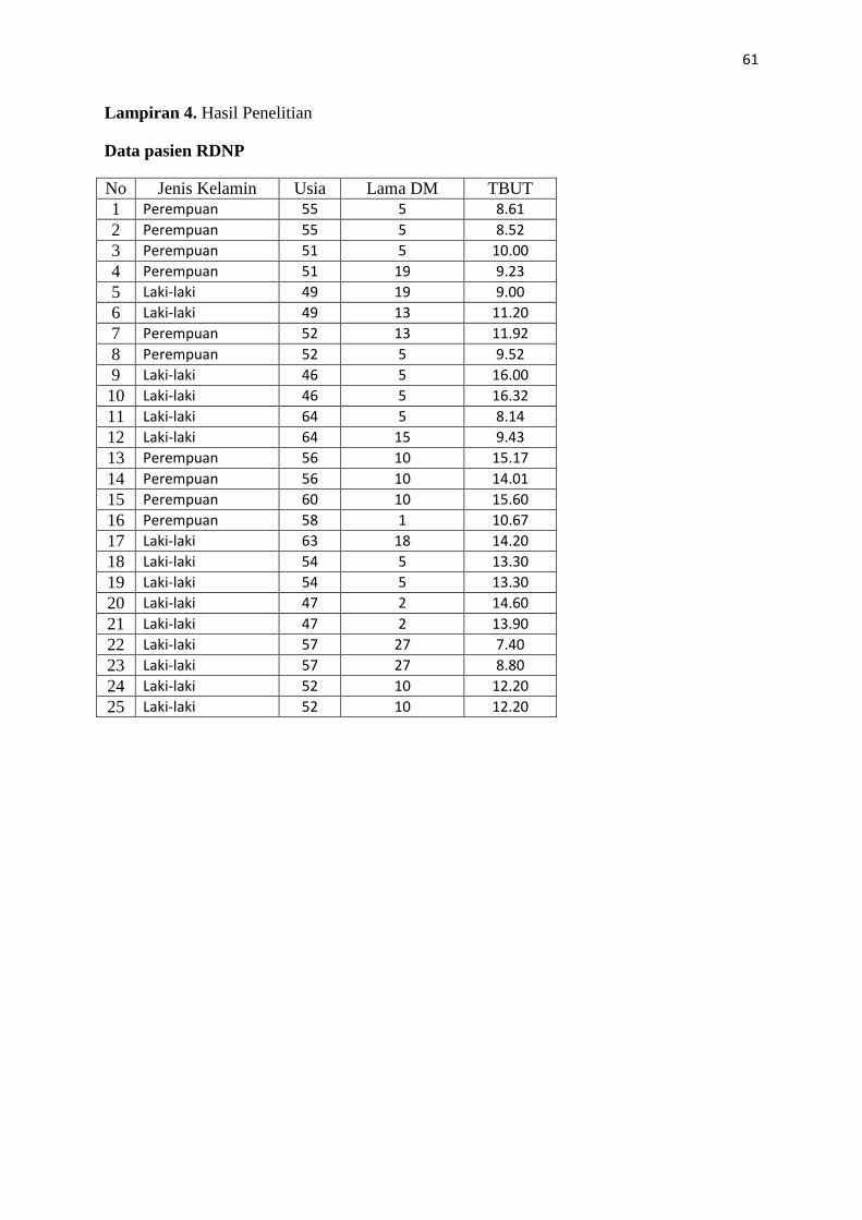
Lampiran 4. Hasil Penelitian
Data pasien RDNP
No Jenis Kelamin Usia Lama DM TBUT
1 Perempuan 55 5 8.61
2 Perempuan 55 5 8.52
3 Perempuan 51 5 10.00
4 Perempuan 51 19 9.23
5 Laki-laki 49 19 9.00
6 Laki-laki 49 13 11.20
7 Perempuan 52 13 11.92
8 Perempuan 52 5 9.52
9 Laki-laki 46 5 16.00
10 Laki-laki 46 5 16.32
11 Laki-laki 64 5 8.14
12 Laki-laki 64 15 9.43
13 Perempuan 56 10 15.17
14 Perempuan 56 10 14.01
15 Perempuan 60 10 15.60
16 Perempuan 58 1 10.67
17 Laki-laki 63 18 14.20
18 Laki-laki 54 5 13.30
19 Laki-laki 54 5 13.30
20 Laki-laki 47 2 14.60
21 Laki-laki 47 2 13.90
22 Laki-laki 57 27 7.40
23 Laki-laki 57 27 8.80
24 Laki-laki 52 10 12.20
25 Laki-laki 52 10 12.20
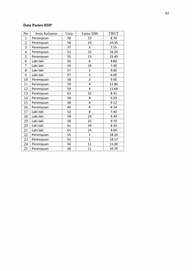
62
Data Pasien RDP
No Jenis Kelamin Usia Lama DM TBUT
1 Perempuan 58 23 8.76
2 Perempuan 58 23 10.35
3 Perempuan 37 3 7.55
4 Perempuan 52 15 16.20
5 Perempuan 55 15 15.40
6 Laki-laki 56 6 9.80
7 Laki-laki 54 19 7.40
8 Laki-laki 57 5 9.00
9 Laki-laki 57 5 6.00
10 Perempuan 58 2 9.00
11 Perempuan 59 4 11.86
12 Perempuan 59 4 11.69
13 Perempuan 62 32 8.35
14 Perempuan 58 8 6.59
15 Perempuan 58 8 9.12
16 Perempuan 46 8 8.14
17 Laki-laki 52 8 7.40
18 Laki-laki 58 25 9.30
19 Laki-laki 58 25 9.70
20 Laki-laki 61 24 8.20
21 Laki-laki 61 24 9.00
22 Perempuan 55 1 18.20
23 Perempuan 55 1 18.17
24 Perempuan 56 11 11.00
25 Perempuan 56 11 10.70
63
Lampiran 5. Hasil Statistik
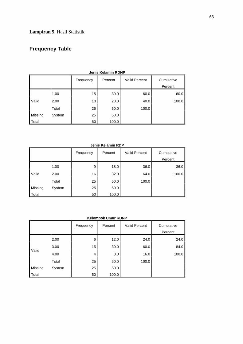
Frequency Table
Jenis Kelamin RDNP
Frequency Percent Valid Percent Cumulative
Percent
Valid
1.00 15 30.0 60.0 60.0
2.00 10 20.0 40.0 100.0
Total 25 50.0 100.0
Missing System 25 50.0
Total 50 100.0
Jenis Kelamin RDP
Frequency Percent Valid Percent Cumulative
Percent
Valid
1.00 9 18.0 36.0 36.0
2.00 16 32.0 64.0 100.0
Total 25 50.0 100.0
Missing System 25 50.0
Total 50 100.0
Kelompok Umur RDNP
Frequency Percent Valid Percent Cumulative
Percent
Valid
2.00 6 12.0 24.0 24.0
3.00 15 30.0 60.0 84.0
4.00 4 8.0 16.0 100.0
Total 25 50.0 100.0
Missing System 25 50.0
Total 50 100.0

64
Kelompok Umur RDP
Frequency Percent Valid Percent Cumulative
Percent
Valid
1.00 1 2.0 4.0 4.0
2.00 1 2.0 4.0 8.0
3.00 20 40.0 80.0 88.0
4.00 3 6.0 12.0 100.0
Total 25 50.0 100.0
Missing System 25 50.0
Total 50 100.0
Kelompok DM RDNP
Frequency Percent Valid Percent Cumulative
Percent
Valid
1.00 12 24.0 48.0 48.0
2.00 5 10.0 20.0 68.0
3.00 3 6.0 12.0 80.0
6.00 5 10.0 20.0 100.0
Total 25 50.0 100.0
Missing System 25 50.0
Total 50 100.0
Kelompok DM RDP
Frequency Percent Valid Percent Cumulative
Percent
Valid
1.00 8 16.0 32.0 32.0
2.00 5 10.0 20.0 52.0
3.00 4 8.0 16.0 68.0
6.00 8 16.0 32.0 100.0
Total 25 50.0 100.0
Missing System 25 50.0
Total 50 100.0
65
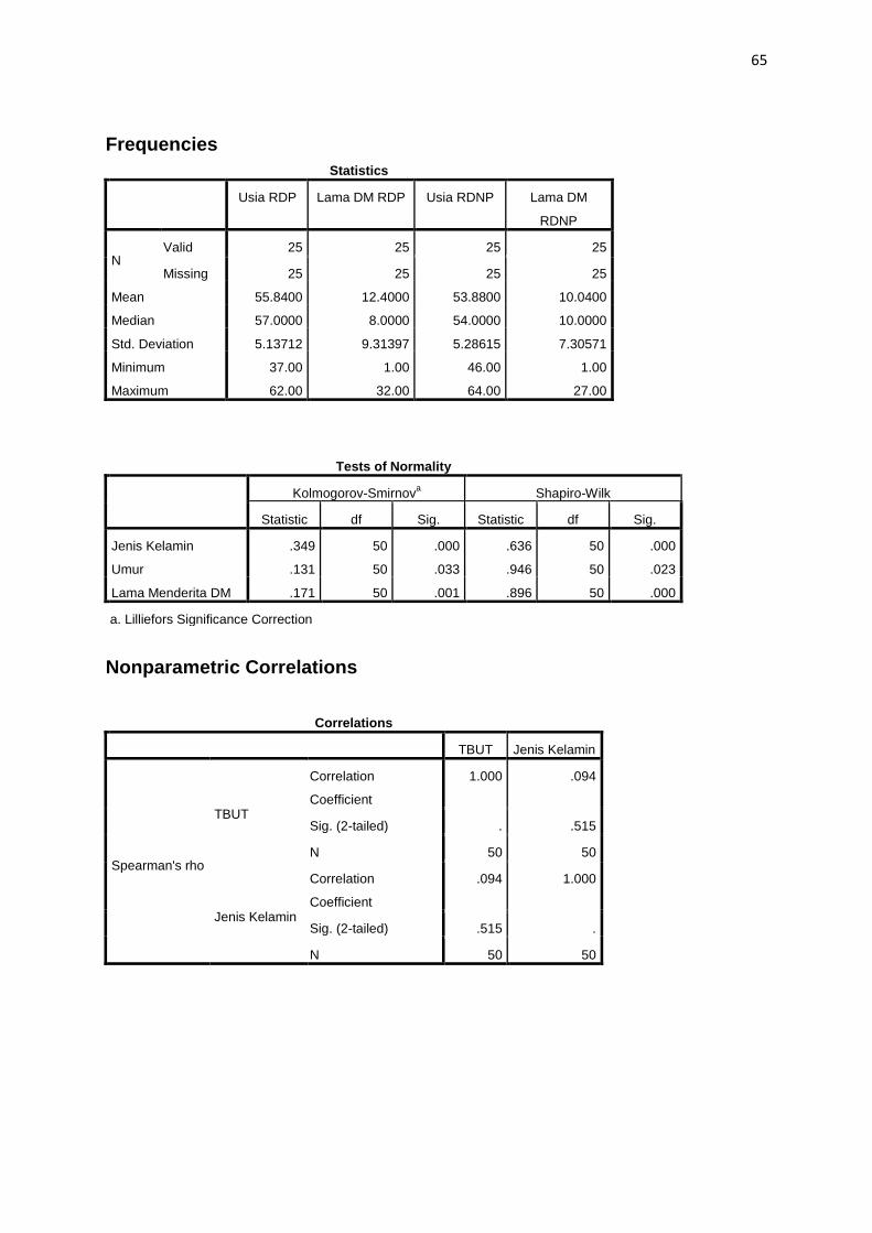
Frequencies
Statistics
Usia RDP Lama DM RDP Usia RDNP Lama DM
RDNP
N Valid 25 25 25 25
Missing 25 25 25 25
Mean 55.8400 12.4000 53.8800 10.0400
Median 57.0000 8.0000 54.0000 10.0000
Std. Deviation 5.13712 9.31397 5.28615 7.30571
Minimum 37.00 1.00 46.00 1.00
Maximum 62.00 32.00 64.00 27.00
Tests of Normality
Kolmogorov-Smirnova Shapiro-Wilk
Statistic df Sig. Statistic df Sig.
Jenis Kelamin .349 50 .000 .636 50 .000
Umur .131 50 .033 .946 50 .023
Lama Menderita DM .171 50 .001 .896 50 .000
a. Lilliefors Significance Correction
Nonparametric Correlations
Correlations
TBUT Jenis Kelamin
Spearman's rho
TBUT
Correlation
Coefficient
1.000 .094
Sig. (2-tailed) . .515
N 50 50
Jenis Kelamin
Correlation
Coefficient
.094 1.000
Sig. (2-tailed) .515 .
N 50 50

66
Nonparametric Correlations
Correlations
Umur TBUT
Spearman's rho
Umur
Correlation Coefficient 1.000 -.232
Sig. (2-tailed) . .105
N 50 50
TBUT
Correlation Coefficient -.232 1.000
Sig. (2-tailed) .105 .
N 50 50
Nonparametric Correlations
Correlations
TBUT Lama
Menderita DM
Spearman's rho
TBUT
Correlation Coefficient 1.000 -.265
Sig. (2-tailed) . .063
N 50 50
Lama Menderita DM
Correlation Coefficient -.265 1.000
Sig. (2-tailed) .063 .
N 50 50
67
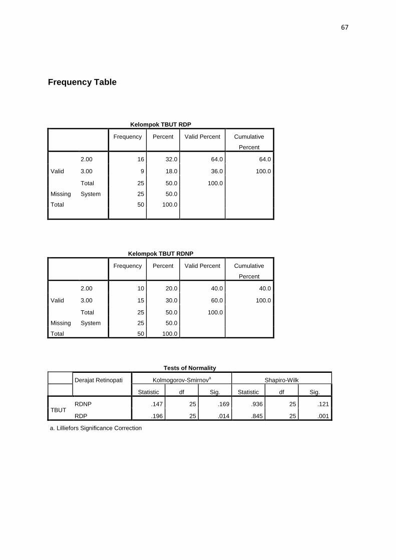
Frequency Table
Kelompok TBUT RDP
Frequency Percent Valid Percent Cumulative
Percent
Valid
2.00 16 32.0 64.0 64.0
3.00 9 18.0 36.0 100.0
Total 25 50.0 100.0
Missing System 25 50.0
Total 50 100.0
Kelompok TBUT RDNP
Frequency Percent Valid Percent Cumulative
Percent
Valid
2.00 10 20.0 40.0 40.0
3.00 15 30.0 60.0 100.0
Total 25 50.0 100.0
Missing System 25 50.0
Total 50 100.0
Tests of Normality
Derajat Retinopati Kolmogorov-Smirnova Shapiro-Wilk
Statistic df Sig. Statistic df Sig.
TBUT RDNP .147 25 .169 .936 25 .121
RDP .196 25 .014 .845 25 .001
a. Lilliefors Significance Correction
68
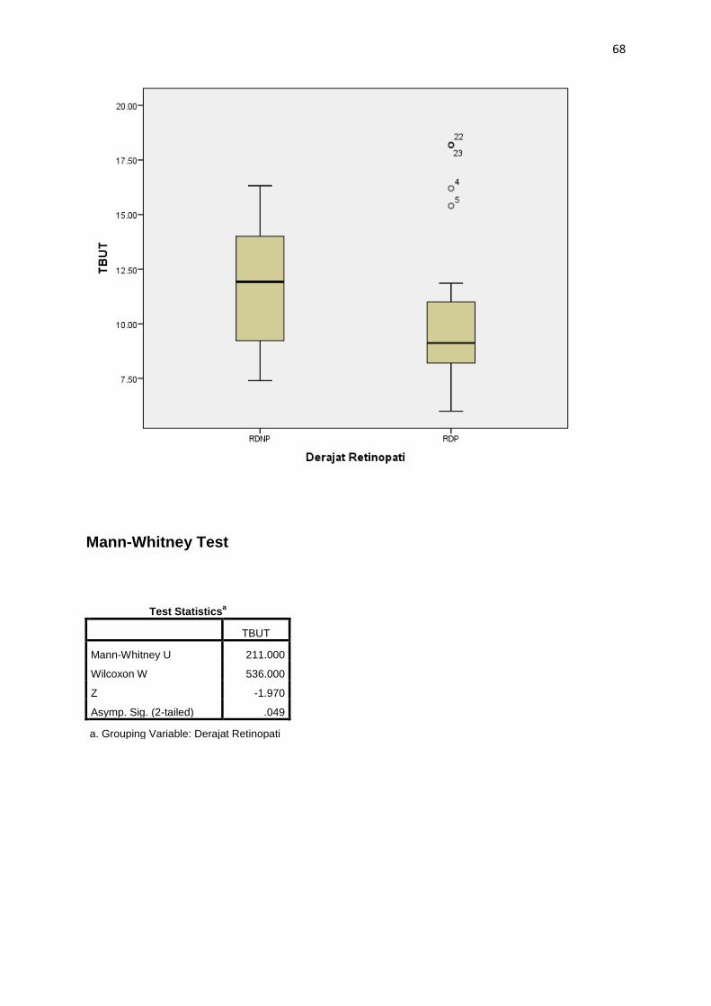
Mann-Whitney Test
Test Statisticsa
TBUT
Mann-Whitney U 211.000
Wilcoxon W 536.000
Z -1.970
Asymp. Sig. (2-tailed) .049
a. Grouping Variable: Derajat Retinopati
69
Lampiran 6. Biodata Mahasiswa
Identitas
Nama : Michelle Abigail
NIM : 22010112130112
Tempat/tanggal lahir : Surabaya / 20 Juni 1995
Jenis Kelamin : Perempuan
Alamat : Jl. Galang Sewu Raya no 1A Baskoro, Tembalang, Semarang
Nomor HP : 085747182653
e-mail : [email protected]
Riwayat Pendidikan Formal
1. SD : SDK Maria Fatima Lulus tahun : 2006
2. SMP : SMPK Ora et Labora Lulus tahun : 2008
3. SMA : SMAK Penabur Gading Serpong Lulus tahun : 2011
4. S1 : Fakultas Kedokteran Universitas Diponegoro Masuk tahun:2012
Keanggotaan Organisasi
1. Steering comitte RHEU Tahun: 2015/2016
2. Ketua Divisi Interna RHEU Tahun : 2014/2015
3. Anggota Divisi Eksterna Tahun : 2013/2014